

Review
doi: 10.1038/ncb0805-750.
Weighing in on ubiquitin: the expanding role of mass-spectrometry-based proteomics
Affiliations
- PMID: 16056266
- PMCID: PMC1224607
- DOI: 10.1038/ncb0805-750
Item in Clipboard
Review
Weighing in on ubiquitin: the expanding role of mass-spectrometry-based proteomics
Nat Cell Biol.
2005 Aug.
Abstract
Mass-spectrometry-based proteomics has become an essential tool for the qualitative and quantitative analysis of cellular systems. The biochemical complexity and functional diversity of the ubiquitin system are well suited to proteomic studies. This review summarizes advances involving the identification of ubiquitinated proteins, the elucidation of ubiquitin-modification sites and the determination of polyubiquitin chain linkages, as well as offering a perspective on the application of emerging technologies for mechanistic and functional studies of protein ubiquitination.
Figures
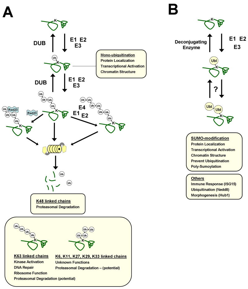
Mechanisms of protein modification by ubiquitin (a) and Ubl proteins (b). (a) Protein substrates are modified by ubiquitin and Ubl-proteins at a lysine (K) residue(s). (1) Ubiquitin attachment to a substrate is catalyzed by the coordinated actions of an E1[dk29 ] activating enzyme, E2 conjugating enzyme, and E3 ligase (2) Poly- ubiquitin chains can be formed through any of 7 lysine residues within ubiquitin. Deubiquitinating enzymes (DUBs) reverse ubiquitination and shorten poly-ubiquitin chains. (3) A subset of factors, termed E4 enzymes, can further lengthen poly- ubiquitin chains. (4, 5) Degradation of ubiquitinated proteins occurs through a mechanism largely dependent on ubiquitin-ubiquitin linkages formed through K48 of ubiquitin. Poly- ubiquitin chain binding proteins such as Rad23/Hhr23 and Dsk2/ Ubiquilin[dk30 ] facilitate recognition and degradation of ubiquitinated substrates by the 26S proteasome. Substrate modifications by mono-ubiquitin, K63-linked chains, or by Ubl-proteins (b) regulate a series of proteasome independent cellular processes. Biological significance of other ubiquitin linkages and many Ubl proteins remain poorly characterized. (b) [dk31 ](1) Ubl attachment occurs through a mechanism analogous to ubiquitination, involving E1, E2, and E3 enzymes. Ubl deconjugating enzymes remove Ubl modifications from substrates. (2) Poly-Ubl chains can be formed for SUMO. Poly-chain formation for other Ubls is currently under investigation. Modification by Ubl proteins can block ubiquitination sites, activate enzymes (including Ub E3 ligases), and affect protein localization.
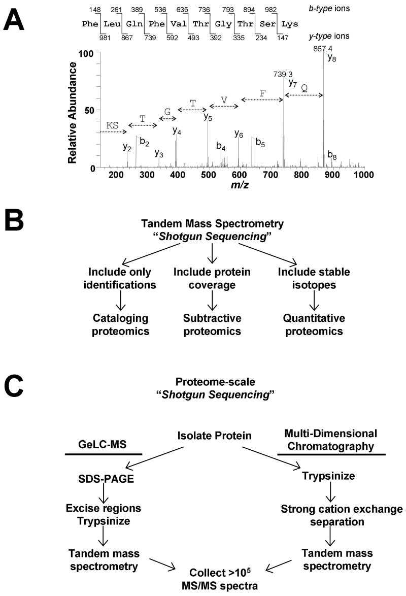
Overview of shotgun sequencing from complex mixtures by mass spectrometry. a) Representative tandem mass (MS/MS) spectrum of a peptide from the protein Urebp1. Amino acid sequence for a single peptide can be deduced from the series of fragment ions present in the spectrum. b) Large-scale peptide detection via shotgun sequencing can be interpreted in three different ways to provide either lists (cataloging proteomics), differential identifications (subtractive proteomics), or abundance comparisons (quantitative proteomics). c) For proteome-scale analyses, huge numbers of MS/MS spectra are collected. Only two routes have proven successful for identifying thousands of proteins from a single sample. Both strategies utilize multiple[dk33 ] steps to fractionate the original sample prior to MS analysis. In one strategy, SDS-PAGE separation followed by tandem MS analysis of many gel regions (sometimes called GeLC-MS) is used. When using multiple-dimensional chromatography, protein mixtures are directly proteolyzed, and the peptide mixture is separated first by strong cation exchange [dk34 ]chromatography. In both cases, the final step involves reversed-phase separation of peptides from multiple samples, followed by tandem MS analysis of multiple samples. Both techniques provide the opportunity to collect hundreds of thousands of MS/MS spectra from a single sample in less than 24 hrs.
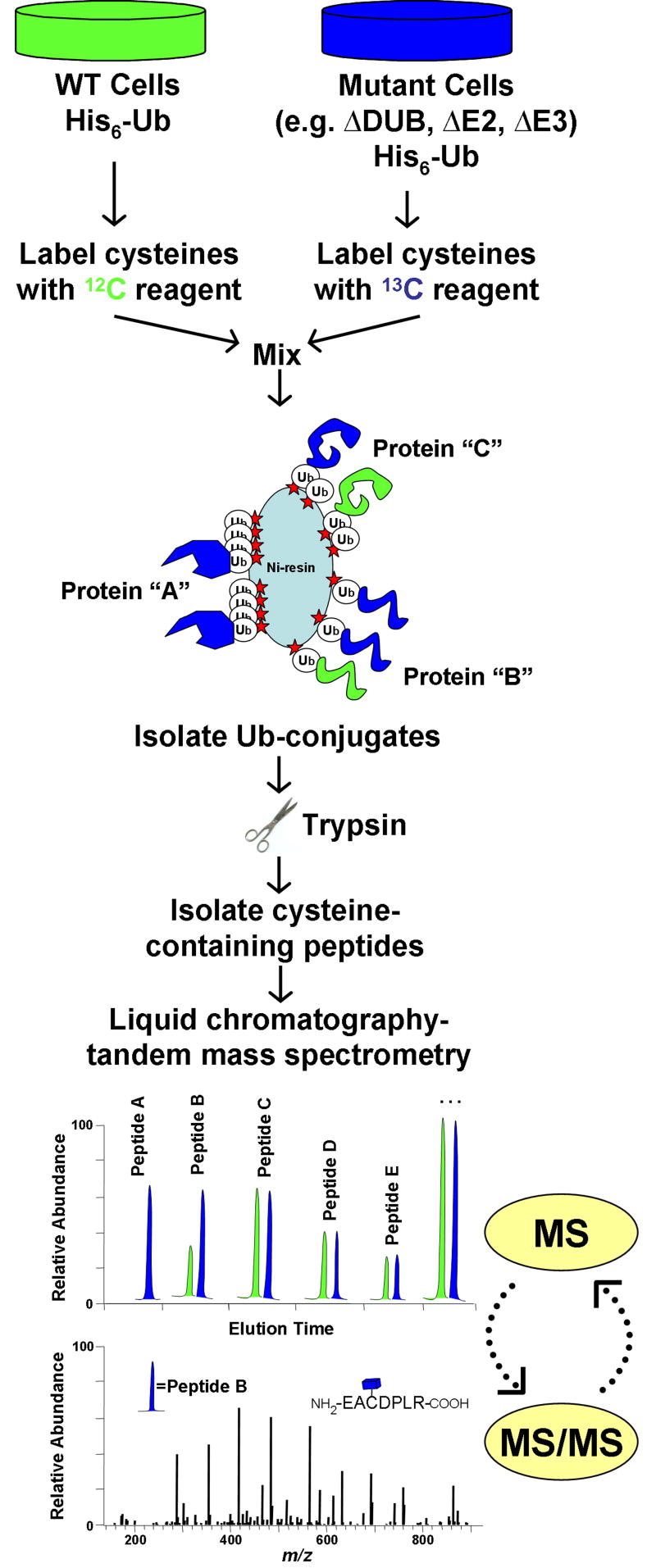
Quantitative profiling of ubiquitin-conjugates using stable isotopes. The isotope coded affinity tag (ICAT) strategy is shown for comparing ubiquitinated proteins between wild type cells and mutant cells lacking a ubiquitin pathway enzyme (e.g. DUB, E2, E3). In an ICAT experiment, protein is harvested from two samples and differentially labeled at cysteinyl residues with either a C- or C-containing reagent. After labeled proteins are mixed, ubiquitinated-proteins are affinity-purified and digested into peptides. The ICAT label allows for further enrichment of cysteine-containing peptides, thus eliminating all peptides derived from ubiquitin. Since C- and C-containing peptides co-elute by reverse-phase chromatography, they can be simultaneously quantified during MS analysis. Rapid cycling between MS and MS/MS modes allows for the acquisition of both sequence and abundance information for isotopic peptide pairs. In this example, protein “A” is exclusively ubiquitinated in mutant cells, while protein “B” is ubiquitination is increased in mutant cells. Protein “C” is ubiquitinated equally in both samples. Other potential strategies would utilize similar work-flows with minor modifications. For example, metabolic labeling (e.g. SILAC) involves incorporation of isotopes into living cells prior to harvest, and would not utilize the cysteine-enrichment step. A benefit of metabolic labeling would be quantification of non-cysteine containing peptides, which would include most –GG signature peptides.
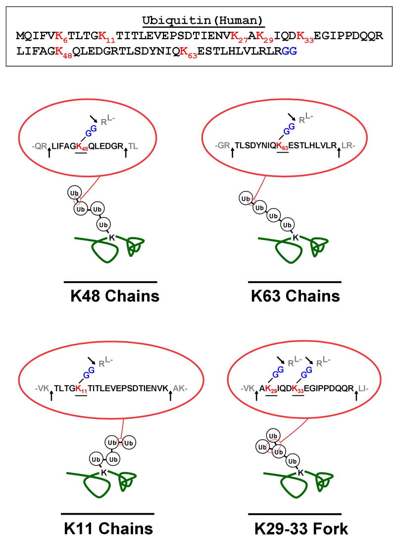
Detecting unique diglycine (–GG) signature peptides for each poly- ubiquitin chain linkage. Each poly-ubiquitin chain conformation can be detected by monitoring a unique signature peptide containing a –GG modified lysine residue, produced by trypsin cleavage. The full amino acid sequence of human ubiquitin is shown at the top. Ubiquitin-ubiquitin linkages correspond to isopeptide bonds formed between the C-terminal glycine (blue) of one ubiquitin and the ɛ-amino group of a lysine residue (red) within the second. These linkages can be formed through any of seven lysine residues (K6, K11, K27, K29, K33, K48, and K63) (red). As an example, K48, K63, and K11 chains are demonstrated. Digestion of these linkages with trypsin produces peptides with distinct amino acid sequences (see insets). Trypsin cleaves at lysine and arginine residues within both primary and branched ubiquitin molecules (see arrows in insets), but cannot cut at lysines modified by isopeptide linked ubiquitin (see underlined in insets). The resulting tryptic peptides contain a –GG modified lysine, bearing an additional mass of 114.04 Da, denoting the original position of the modification. Database searching algorithms can utilize both the missed cleavage and –GG modification as search criteria when assigning precise sites of ubiquitination. In the case of a forked poly-ubiquitin chain, as demonstrated through K29 and K33, two –GG modified lysines are detected on the same peptide.
References
-
- Hershko A, Ciechanover A. The ubiquitin system. Annu Rev Biochem. 1998;67:425–479. - PubMed
-
- Pickart CM. Mechanisms underlying Ubiquitination. Nat Rev Cell Mol Biol. 2001;70:503–533. - PubMed
-
- Finley D, Ciechanover A, Varshavsky A. Ubiquitin as a central cellular regulator. Cell. 2004;116:S29–32. - PubMed
Publication types
MeSH terms
Substances
Grants and funding
LinkOut - more resources
Full Text Sources
Other Literature Sources
