

Effects of salmon calcitonin on trabecular microarchitecture as determined by magnetic resonance imaging: results from the QUEST study
- PMID: 16059627
- PMCID: PMC4445726
- DOI: 10.1359/JBMR.050411
Effects of salmon calcitonin on trabecular microarchitecture as determined by magnetic resonance imaging: results from the QUEST study
Abstract
The unique noninvasive MRI technique was used to assess trabecular microarchitecture at multiple skeletal sites in 91 postmenopausal osteoporotic women receiving nasal spray salmon calcitonin (CT-NS) or placebo over 2 years. In the distal radius and lower trochanter of the hip, individuals treated with CT-NS exhibited significant preservation of trabecular bone microarchitecture compared with placebo, where significant deterioration was shown. MRI analyses of os calcis or microCT/histomorphometric analyses of bone biopsies did not reveal consistent differences in architecture between CT-NS and placebo.
Introduction: It is postulated that the reduction in osteoporotic fracture risk in response to certain antiresorptive osteoporosis therapies is caused less by effects on bone quantity than on bone quality (specifically trabecular microarchitecture). To test this hypothesis, the QUEST study was conducted to assess the effects of nasal spray salmon calcitonin (CT-NS) or placebo on parameters of trabecular microarchitecture at multiple skeletal sites using noninvasive MRI technology and iliac crest bone biopsies by microCT/histomorphometry.
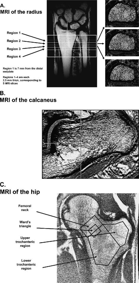
Materials and methods: Ninety-one postmenopausal osteoporotic women were followed for 2 years (n = 46 for CT-NS, n = 45 for placebo); all women received 500 mg calcium daily. MRI measurements at distal radius, hip (T2 relaxation time [T2*]), and os calcis (obtained yearly), iliac crest bone biopsies with 2D histomorphometry and 3D microCT (obtained at study onset and conclusion), DXA-BMD at spine/hip/wrist/os calcis (obtained yearly), and markers of bone turnover (obtained at 2-week to 12-month intervals) were analyzed, with an analysis of covariance model used to assess treatment effect for parameters of interest.
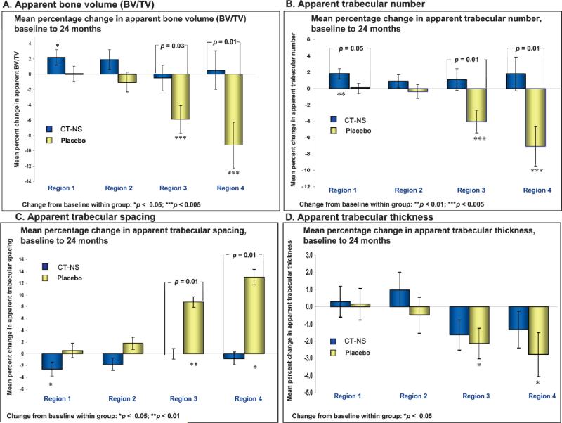
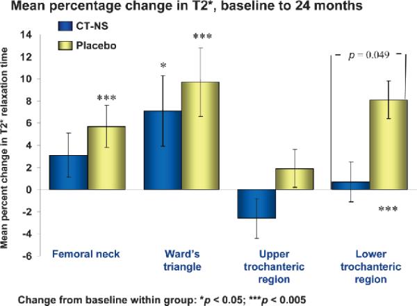
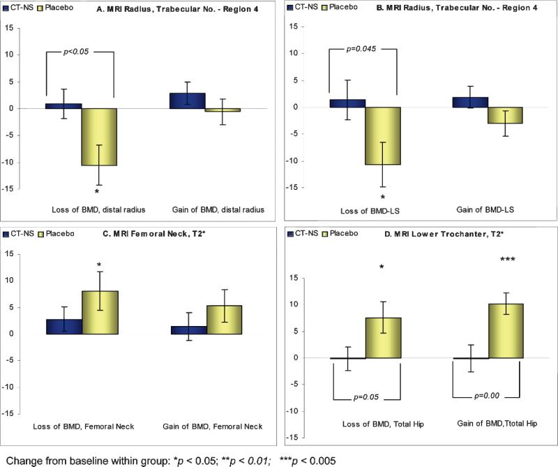
Results and conclusions: MRI assessment of trabecular microarchitecture at individual regions of the distal radius revealed significant improvement, or preservation (no significant loss), in the CT-NS-treated group compared with significant deterioration in the placebo control group, as reflected in apparent BV/TV (p < 0.03), apparent trabecular number (p < 0.01), and apparent trabecular spacing (p < 0.01). Also, at the hip, the CT-NS group exhibited preservation of trabecular microarchitecture at the lower trochanter (p < 0.05) as determined by T2* MRI technology. Significant deterioration of trabecular bone architecture was noted in the placebo group at the femoral neck, Ward's triangle, and lower trochanteric sites. Apart from a significant increase in apparent trabecular number in the CT-NS group, significant changes within or between groups were not noted at the os calcis. Combined microCT/histomorphometric analysis of iliac crest bone biopsies did not reveal significant differences between treated and placebo groups. In the CT-NS group, regardless of the change in BMD (gain or loss) at the spine, hip, or distal radius, preservation of parameters of trabecular microarchitecture was noted, whereas in the placebo group, regardless of the change in BMD (gain or loss) at the spine, hip, or distal radius, loss or preservation was noted; however, changes in DXA/BMD (of the spine, hip, wrist, os calcis) between CT-NS and placebo groups were not significant. Serum C-telopeptide (S-CTx), a specific bone resorption marker, was reduced by 22.5% at 24 months (p = 0.056). The results of the QUEST study suggest therapeutic benefit of CT-NS compared with placebo in maintaining trabecular microarchitecture at multiple skeletal sites and support the use of MRI technology for assessment of trabecular microarchitecture in clinical research trials. However, the results also highlight site specific differences in response to antiresorptive therapies and the importance of sufficiently large sampling volumes (areas) to obtain reliable assessment of bone architecture.
Figures
Similar articles
-
Effect of intermittent administration of 200 IU intranasal salmon calcitonin and low doses of 1alpha(OH) vitamin D3 on bone mineral density of the lumbar spine and hip region and biochemical bone markers in women with postmenopausal osteoporosis: a pilot study.Clin Rheumatol. 2005 Jun;24(3):232-8. doi: 10.1007/s10067-004-1004-6. Epub 2005 Jan 13. Clin Rheumatol. 2005. PMID: 15647969 Clinical Trial.
-
Increases in BMD correlate with improvements in bone microarchitecture with teriparatide treatment in postmenopausal women with osteoporosis.J Bone Miner Res. 2007 Aug;22(8):1173-80. doi: 10.1359/jbmr.070413. J Bone Miner Res. 2007. PMID: 17451369
-
Skeletal effects of cyclic recombinant human growth hormone and salmon calcitonin in osteopenic postmenopausal women.J Clin Endocrinol Metab. 1997 Apr;82(4):1111-7. doi: 10.1210/jcem.82.4.3901. J Clin Endocrinol Metab. 1997. PMID: 9100582 Clinical Trial.
-
Calcitonin for the long-term prevention and treatment of postmenopausal osteoporosis.Bone. 2002 May;30(5 Suppl):75S-79S. doi: 10.1016/s8756-3282(02)00715-9. Bone. 2002. PMID: 12008163 Review.
-
Effects of osteoporosis medications on bone quality.Joint Bone Spine. 2007 Jan;74(1):39-47. doi: 10.1016/j.jbspin.2006.06.004. Epub 2006 Nov 28. Joint Bone Spine. 2007. PMID: 17196423 Review.
Cited by
-
Nasal salmon calcitonin blunts bone microstructure alterations in healthy postmenopausal women.Osteoporos Int. 2015 Jan;26(1):383-93. doi: 10.1007/s00198-014-2937-5. Epub 2014 Oct 22. Osteoporos Int. 2015. PMID: 25566730 Clinical Trial.
-
A longitudinal HR-pQCT study of alendronate treatment in postmenopausal women with low bone density: Relations among density, cortical and trabecular microarchitecture, biomechanics, and bone turnover.J Bone Miner Res. 2010 Dec;25(12):2558-71. doi: 10.1002/jbmr.157. Epub 2010 Jun 18. J Bone Miner Res. 2010. PMID: 20564242 Free PMC article. Clinical Trial.
-
Clinical perspectives on bone quality in osteoporosis: effects of drug therapy.Drugs Aging. 2007;24(7):529-35. doi: 10.2165/00002512-200724070-00001. Drugs Aging. 2007. PMID: 17658904 Review.
-
Comparing and contrasting the effects of strontium ranelate and other osteoporosis drugs on microarchitecture.Osteoporos Int. 2010 Jun;21 Suppl 2:S437-42. doi: 10.1007/s00198-010-1250-1. Epub 2010 May 13. Osteoporos Int. 2010. PMID: 20464378 Review.
-
Implications of resolution and noise for in vivo micro-MRI of trabecular bone.Med Phys. 2008 Dec;35(12):5584-94. doi: 10.1118/1.3005598. Med Phys. 2008. PMID: 19175116 Free PMC article.
References
-
- National Institutes of Health (NIH) Consensus Development Panel 2000 Osteoporosis Prevention, Diagnosis, and Therapy. NIH Consensus Statement. 2001 Available online at http://consensus.nih.gov. Accessed.
-
- Chesnut CH, Rosen CJ. Reconsidering the effects of antiresorptive therapies in reducing osteoporotic fracture (Perspective). J Bone Miner Res. 2001;16:2163–2172. - PubMed
-
- Delmas PD. How does antiresorptive therapy decrease the risk of fracture in women with osteoporosis? Bone. 2000;27:1–3. - PubMed
-
- Riggs BL, Melton LJ. Bone turnover matters: The raloxofene paradox of dramatic decreases in vertebral fractures without commensurate increases in bone density. J Bone Miner Res. 2002;17:11–14. - PubMed
-
- Chesnut CH, Silverman S, Andriano K, Genant HK, Gimona A, Harris S, Kiel D, LeBoff M, Maricic M, Miller P, Moniz C, Peacock M, Richardson P, Watts N, Baylink D. A randomized trial of nasal spray salmon calcitonin in postmenopausal women with established osteoporosis: The Prevent Recurrence of Osteoporotic Fracture study. Am J Med. 2000;109:267–276. - PubMed
Publication types
MeSH terms
Substances
Grants and funding
LinkOut - more resources
Full Text Sources
Medical
Miscellaneous
