

Oocyte maturation: the coming of age of a germ cell
- PMID: 16059829
- PMCID: PMC1482430
- DOI: 10.1055/s-2005-872451
Oocyte maturation: the coming of age of a germ cell
Abstract
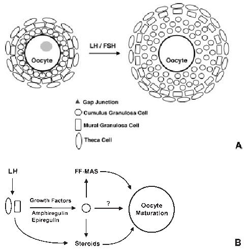
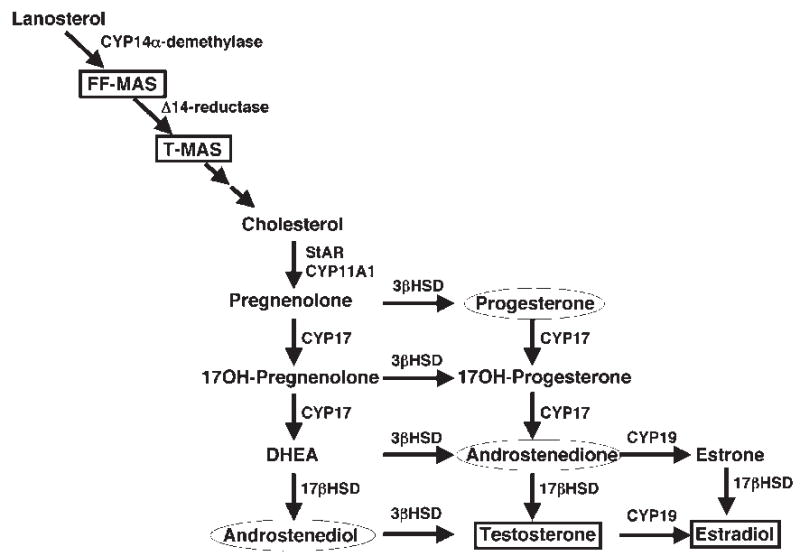
Normal female fertility relies on proper development of the oocyte. This growth culminates just prior to ovulation, when oocyte maturation occurs. Oocyte maturation refers to a release of meiotic arrest that allows oocytes to advance from prophase I to metaphase II of meiosis. This precisely regulated meiotic progression is essential for normal ovulation and subsequent fertilization, and involves changes in the delicate balance between factors promoting meiotic arrest and others that are stimulating maturation. Most of the inhibitory mechanisms appear to involve the upregulation of intracellular cyclic adenosine monophosphate levels. These processes may include direct transport of the nucleotide into oocytes via gap junctions, G protein-mediated stimulation of adenylyl cyclase, and inhibition of intracellular phosphodiesterases. In contrast, potential factors that play roles in triggering oocyte maturation include gonadotropins (e.g., follicle-stimulating factor and luteinizing hormone), growth factors (e.g., amphiregulin and epiregulin), sterols (e.g., follicular fluid-derived meiosis-activating sterol), and steroids (e.g., testosterone progesterone, and estradiol). Delineating the complex interactions between these positive and negative components is critical for determining the role that oocyte maturation plays in regulating follicle development and ovulation, and may lead to novel methods that can be used to modulate these processes in women with both normal and aberrant fertility.
Figures
References
-
- Albertini DF, Carabatsos MJ. Comparative aspects of meiotic cell cycle control in mammals. J Mol Med. 1998;76:795–799. - PubMed
-
- Albertini DF, Combelles CM, Benecchi E, Carabatsos MJ. Cellular basis for paracrine regulation of ovarian follicle development. Reproduction. 2001;121:647–653. - PubMed
-
- diZerega GS, Hodgen GD. Folliculogenesis in the primate ovarian cycle. Endocr Rev. 1981;2:27–49. - PubMed
-
- Griffin JE, Ojeda SR. Textbook of Endocrine Physiology, 4th ed. New York: Oxford University Press; 2000
-
- Mandl AM, Zuckerman S. The relation of age to numbers of oocytes. J Endocrinol. 1951;7:190–193. - PubMed
Publication types
MeSH terms
Substances
Grants and funding
LinkOut - more resources
Full Text Sources
Other Literature Sources
Miscellaneous
