Review
. 2005;7(1):17-29.
doi: 10.31887/DCNS.2005.7.1/frreedman.

Early biomarkers of psychosis

Affiliations
Review

Early biomarkers of psychosis

Robert Freedman et al. Dialogues Clin Neurosci. 2005.

Abstract

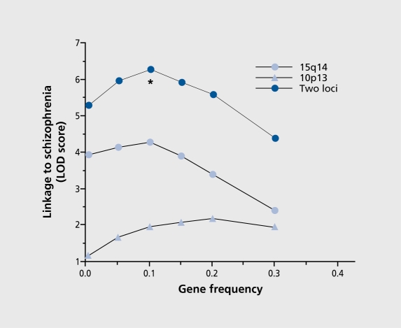
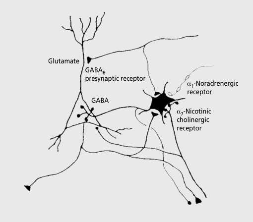
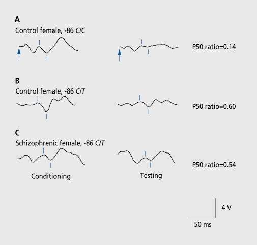
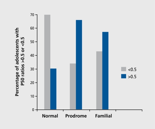
Biological traits that are predictive of the later development of psychosis have not yet been identified. The complex, multidetermined nature of schizophrenia and other psychoses makes it unlikely that any single biomarker will be both sensitive and specific enough to unambiguously identify individuals who will later become psychotic. However, current genetic research has begun to identify genes associated with schizophrenia, some of which have phenotypes that appear early in life. While these phenotypes have low predictive power for identifying individuals who will become psychotic, they do serve as biomarkers for pathophysiological processes that can become the targets of prevention strategies. Examples are given from work on the role of the alpha(T)nicotinic receptor and its gene CHRNA7 on chromosome 15 in the neurobiology and genetic transmission of schizophrenia.

Aun no se han identificado los rasgos biológicos que predicen el desarrollo posterior de una psicosis. La naturaleza compleja y multideterminada de la esquizofrenia y de otras psicosis hace poco probable que algún biomarcador aislado sea lo suficientemente sensible y aspecífico para identificar con certeza sujetos que más tarde llegarán a ser psicóticos. Sin embargo, la investigación genética actual ha comenzado a identificar genes que se asocian con la esquizofrenia, algunos de los cuales tienen fenotipos qua aparacen precozmente en la vida. Aunque estos fenotipos tienen un bajo podar predictor para identificar individuos qua llegarán a ser psicóticos, ellos sirven como biomarcadores de procesos fisiopatológicos que pueden llegar a ser los blancos da las estrategias da prevención. Ejemplos de esto provienen del trabajo acerca del papel del receptor nicotínico α7 y su gen CHRNA 7 en el cromosoma 15 en la neurobiología y en la transmisión genética de la esquizofrenia.

Les particularités biologiques prédictives du développement ultérieur d'une psychose n'ont pas encore été identifiées. La nature complexe, multi-factorielle de la schizophrénie et d'autres psychoses rend peu probable qu'un seul biomarqueur soit a la fois suffisamment sensible et spécifique pour permettre d'identifier sans ambiguïté les individus qui deviendront psychotiques. Cependant, la recherche génétique actuelle a commence a identifier des gènes associes a la schizophrénie, certains ayant des phénotypes qui apparaissent précocement dans le cours de la vie. Alors que ces phénotypes ont un faible pouvoir prédictif pour identifier les individus qui développeront une psychose, ils servent de biomarqueurs pour les processus physiopathologiques pouvant devenir les cibles des stratégies de prévention. Des exemples sont fournis par un travail sur le rôle du récepteur α7-nicotinique et de son gène CHRNA7 situe sur le chromosome 15, en neurobiologie et dans la transmission génétique de la schizophrénie.


Figures

Figure 1.
Figure 1.. Relationship of maximum LOD score to gene frequencies for two locus linkage to schizophrenia. This model produces a peak LOD score at allele frequencies of 0.1 , which is significantly greater than the score derived from the heterogeneous model (Chisquare=14.54; df=1; P=0.000069).
Reproduced from reference 2: Freedman R, Leonard S, Olincy A, et al. Evidence for the multigenic inheritance of schizophrenia. Am J Med Genet. 2001;105:794-800. Copyright © 2001, Wiley-Liss, Inc.
Figure 2.
Figure 2.. Neurophysiological studies of the P50 sensory gating phenomena have implicated the pyramidal neurons of the hippocampus as a source of the wave. Interneurons in the hippocampus are responsible for inhibition of pyramidal neuron response in the conditioning-testing or paired stimulus paradigm. These interneurons are excited by cholinergic innervation via α7-nicotinic cholinergic receptors and inhibited by adrenergic receptors. GABA, γ-aminobutyric acid.
Figure 3.
Figure 3.. Effects of a single nucleotide polymorphism in the promoter of CHRNA7 on P50 sensory gating. The normal subject with more common alleles (-86 C/C) has normal suppression of P50, measured as a low P50 test to conditioning amplitude ratio. The subjects with the polymorphism (-86 C/T) have higher, more abnormal ratios. The physiological abnormality is expressed regardless of whether the subject is normal or has schizophrenia.
Reproduced from reference 9: Leonard S, Gault J, Hopkins J, et al. Association of promoter variants in the α7-nicotinic acetylcholine receptor subunit gene with an inhibitory deficit found in schizophrenia. Arch Gen Psychiatry. 2002;59:1085-1096. Copyright © 2002, American Medical Association.
Figure 4.
Figure 4.. Percentages of P50 ratios in the abnormal range (amplitude of test wave/amplitude of conditioning wave>0.5) is elevated in adolescents with prodromal symptoms of schizophrenia or with first-degree relatives who have schizophrenia. Data are from a study conducted on the island Palau by Myles-Worsley etal.
Figure 5.
Figure 5.. Relationship between age and selected electrophysiological variables. A. P50 T/C ratio generally decreases (ie, sensory gating improves) with advancing age. B. Electroencephalogram (EEG) spectral power in the θ band (4-8 Hz), associated with rapid-eye movement (REM) sleep in the hippocampus, also increases in the same time period.
Reproduced from reference 12: Kisley MA, Olincy A, Robbins E, et al. Sensory gating impairment associated with schizophrenia persists into REM sleep. Psychophysiology. 2003;40:29-38. Copyright © 2003, Blackwell Publishing.
Figure 6.
Figure 6.. In schizophrenia, there are several abnormalities in smooth pursuit eye movements. One of these is elevated frequency of anticipatory saccades, the failure to inhibit fast or saccadic eye movements that cause the eyes to jump ahead of the slowly moving target. Broken line, target position; unbroken line, eye position.
Figure 7.
Figure 7.. Functional magnetic resonance imaging of 14 schizophrenics and 14 normals performing smooth pursuit eye movements. The images are comparisons between the two groups. The schizophrenics have increased hemodynamic activity in the hippocampus, consistent with loss of inhibition.
Reproduced from reference 14: Tregellas JR, Tanabe JL, Miller DE, Ross RG, Olincy A, Freedman R. Neurobiology of smooth pursuit eye movement deficits in schizophrenia: an fMRI study. Am J Psychiatry. 2004;161:315-321 . Copyright © 2004, American Psychiatric Association.
Figure 8.
Figure 8.. Admixture analysis of the frequency (per minute) of leading saccades during a smooth-pursuit eye movement task in 189 children, aged 6 to 15 years. For the typically developing children, 81% perform in the lowest (best-performing) mode and 19% in the moderately elevated mode. First-degree relatives of persons with schizophrenia are equally distributed, with 50% in the lowest mode and 50% in the moderately elevated mode. For children who have schizophrenia, 6% have leading saccade frequencies in the lowest mode, 86% in the moderately elevated mode, and 8% in the severely elevated mode.
Reproduced from reference 16: Ross RG. Early expression of a pathophysiological feature of schizophrenia: saccadic intrusions into smooth-pursuit eye movements in school-age children vulnerable to schizophrenia. J Am Acad Child Adolescent Psychiatry. 2003;42:468-476. Copyright © 2003, Lippincott, Williams and Wilkins.
Figure 9.
Figure 9.. Dual labeling with an antibody for the inhibitory neurotransmitter GABA (left) and radioactive α-bungarotoxin (right), which labels α7-nicotinic receptors, demonstrates expression of these receptors on inhibitory interneurons in the rat hippocampus. For other examples, see reference 1. GABA, γ-aminobutyric acid.
Figure 10.
Figure 10.. Expression of α7-nicotinic receptors in the hippocampus, measured by labeling with α-bungarotoxin (α-BTX), in rats during early development and adulthood.
Reproduced from reference 23: Adams CE, Stitzel JA, Collins AC, Freedman R. α7-Nicotinic receptor expression and the anatomical organization of hippocampal interneurons. Brain Res. 2001;922:180-190. Copyright © 2001, Elsevier.
Figure 11.
Figure 11.. Developmental time course of α7-nicotinic receptors in DBA/2 mice (right) and a control strain, C3H (left). Prenatal days 13, 16, 17, and 18 are shown from top to bottom. A. Binding for α-bungarotoxin (α-BTX) is initially observed in the dorsal portion of the hippocampal anlage of C3H mice on embryonic day 13 (E13). Binding is both diffuse (arrowheads) and clumped (arrows) in appearance. No α-BTX binding is observed in the neuroepithelium (NE). B. Binding of α-BTX is not detectable in the developing hippocampus of DBA/2 mice on E13. C. Binding of α-BTX is increased in density and is present in both hippocampal areas CA1 and CA3 of the C3H mouse strain on E16, but not in the immature dentate gyrus (D). The pattern of hippocampal α-BTX binding is again comprised of a mixture of diffuse (arrowheads) and clumped (arrows) silver grains. D. Binding of α-BTX is initially detected in the hippocampal formation of DBA/2 mice on E16. The level of binding is higher in area CA3 than in area CA1 , while no binding is observed in the dentate gyrus. Scattered clumps of silver grains (arrows) are seen within a more diffuse background (arrowheads). Overall, the level of α-BTX binding in DBA/2 mouse hippocampus on E16 is much lower than that observed in C3H mouse hippocampus at the same age. E. Binding of α-BTX is increased in density in hippocampal areas CA1 and CA3 and is initially detected in the dentate gyrus (D) on E17 in C3H mice (arrowheads). F. The level of α-BTX binding is also increased in hippocampal areas CA1 and CA3 in DBA/2 mice on E17, but is not detectable in the dentate gyrus. G. A marked increase in the density of α-BTX binding is seen in hippocampal area CA1 of C3H mice on E18, especially at the border between stratum pyramidale and stratum radiatum of area CA1 (arrows). α-BTX binding density is also increased in hippocampal area CA3 and in the dentate gyrus of C3H mice at this stage of development. H. Binding of α-BTX is initially observed (arrowheads) in the dentate gyrus (D) of DBA/2 mice on E18. An increase in the density of α-BTX binding is also seen in both hippocampal areas CA1 and CA3 of DBA/2 mice at this age, although binding levels are still greater in area CA3 than in area CA1 . Calibration bar =120 mm.
Reproduced from reference 24: Adams CE. Comparison of α7-nicotinic acetylcholine receptor development in the hippocampal formation of C3H and DBA/2 mice. Brain Res Dev Brain Res. 2003;143:137-149. Copyright © 2003, Elsevier.

References

    1. Freedman R. N Engl J Med. 2003;349:1738–1749. - PubMed
    1. Freedman R., Leonard S., Olincy A., et al. Evidence for the multigenic inheritance of schizophrenia. Am J Med Genet. 2001;105:794–800. - PubMed
    1. Brzustowicz LM., Hodgkinson KA., Chow EW., Honer WG., Bassett AS. Location of a major susceptibility locus for familial schizophrenia on chromosome 1q21-q22. Science. 2000;288:678–682. - PMC - PubMed
    1. Pulver AE., Lasseter VK., Kasch L., et al. Schizophrenia: a genome scan targets chromosomes 3p and 8p as potential sites of susceptibility genes. Am J Med Genet. 1995;60:252–260. - PubMed
    1. Freedman R., Coon H., Myles-Worsley M., et al. Linkage of a neurophysiology deficit in schizophrenia to a chromosome 15 locus. Proc Natl Acad Sci USA. 1997;94:587–592. - PMC - PubMed

Publication types

MeSH terms