. 2005 Aug 1;170(3):401-11.
doi: 10.1083/jcb.200501138.

The limited role of NH2-terminal c-Jun phosphorylation in neuronal apoptosis: identification of the nuclear pore complex as a potential target of the JNK pathway

Affiliations

The limited role of NH2-terminal c-Jun phosphorylation in neuronal apoptosis: identification of the nuclear pore complex as a potential target of the JNK pathway

Cagri G Besirli et al. J Cell Biol. .

Abstract

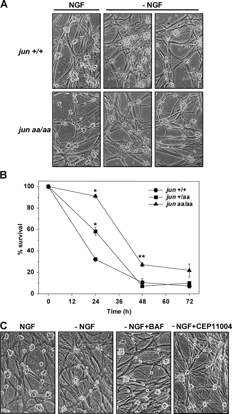
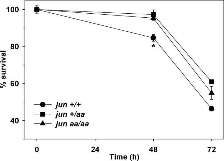
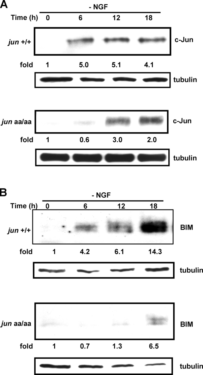
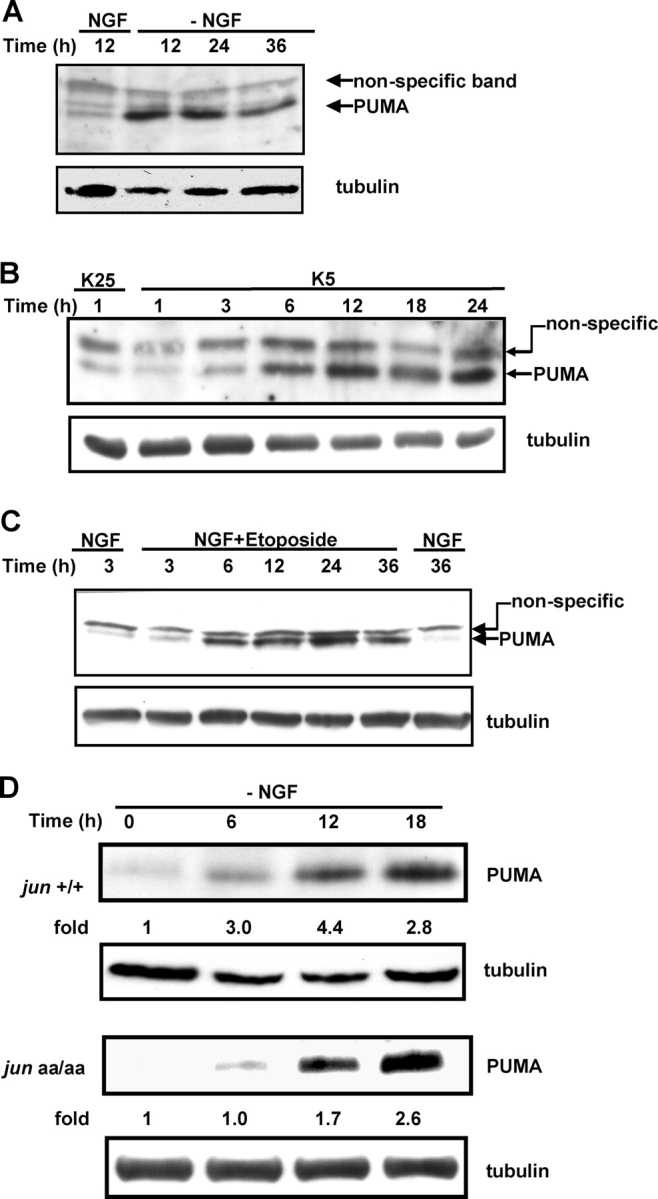
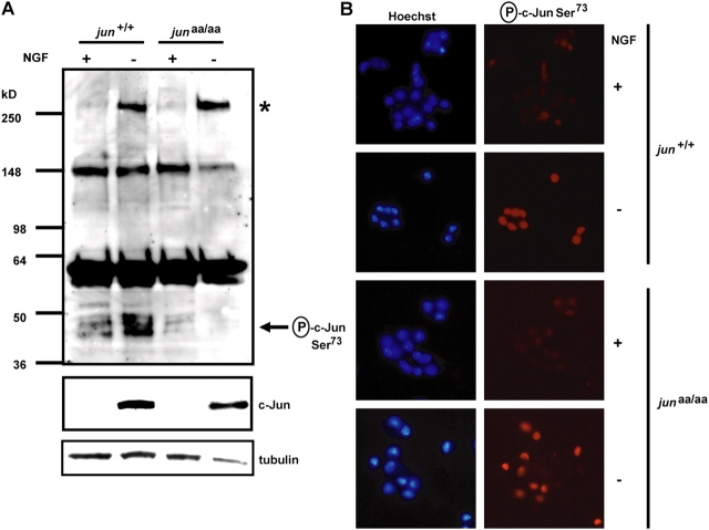
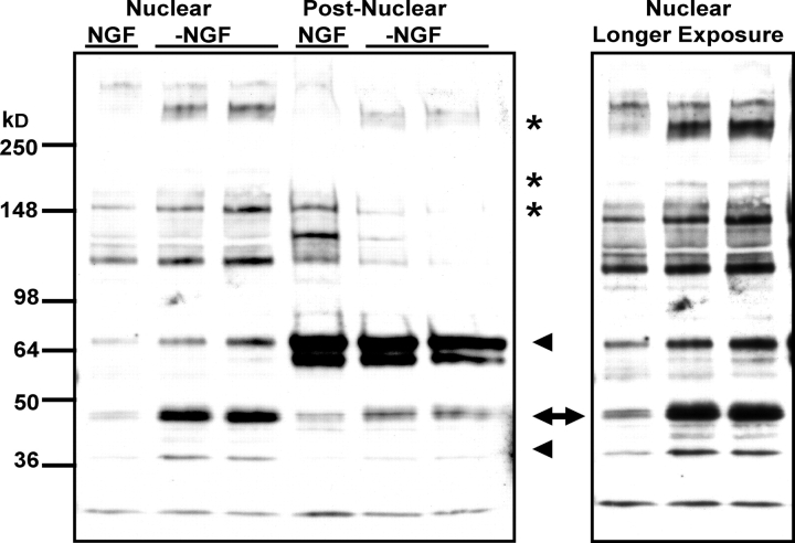
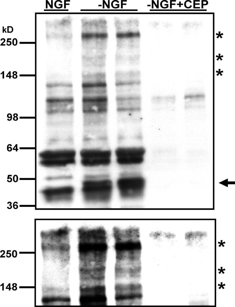
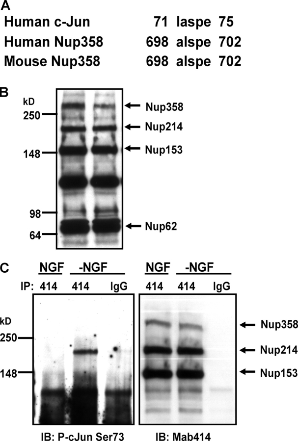
c-Jun is induced in many neuronal death paradigms. A critical step in c-Jun regulation involves phosphorylation of Ser63/Ser73 located in the NH2-terminal transactivation domain. To determine the importance of this phosphorylation for neuronal apoptosis, we analyzed the sympathetic neurons of mice carrying a mutant c-Jun gene that lacks Ser63/Ser73 phosphorylation sites (jun aa). Trophic factor-deprivation or DNA damage-induced death was significantly delayed in jun aa/aa neurons. Neuronal c-Jun induction was only partially inhibited, demonstrating that phosphorylation of Ser63/73 is not required for c-Jun activation. The inductions of proapoptotic BH3-only proteins, Bim and PUMA/Bbc3, were delayed during neuronal apoptosis in mutant neurons. These results demonstrate that NH2-terminal c-Jun phosphorylation is important, but not necessary, for the induction of proapoptotic genes and neuronal apoptosis. Thus, additional JNK substrates may be critical for neuronal death. As potential mediators, we identified additional nuclear MLK/JNK substrates, including Nup214 subunit of the nuclear pore complex.


Figures

Figure 1.
Figure 1.
Lack of c-Jun phosphorylation increases the number of sympathetic neurons isolated from the SCG. Sympathetic neurons were isolated from the SCGs of newborn littermate mice and grown in NGF-containing medium. (A) Phase-contrast images of sympathetic neurons from two different genotypes. Bar, 40 μm. (B) Total number of sympathetic neurons was determined by counting viable neurons after 6–12 d in culture. Mean of wild-type neurons was set to 100%. More sympathetic neurons were isolated from the SCGs of mutant mice compared with wild-type littermates (*, P < 0.01, t test). Data are represented as mean ± SE from five independent experiments with 4–10 mice.
Figure 2.
Figure 2.
Lack of NH2-terminal c-Jun phosphorylation impairs trophic factor deprivation–induced apoptosis in sympathetic neurons. (A) Phase-contrast images of 5-DIV–sympathetic neurons deprived of NGF (−NGF) for 33 h. Bar, 40 μm. (B) Sympathetic neurons isolated from littermate mice were deprived of NGF, followed by NGF replacement every 24 h for an additional 5–7 d. Cells were fixed after this rescue period and the number of surviving neurons was determined by counting the cells after crystal violet staining. *, P ≤ 0.001, **, P < 0.01, t test, all compared with wild type. Data are represented as mean ± SE from three independent experiments. (C) Sympathetic neurons were isolated from newborn jun aa/aa mice. Medium with no NGF was provided at 5 DIV (−NGF) for 72 h. The medium for sister cultures contained 1.33 μM of selective MLK-inhibitor CEP11004 (−NGF+CEP11004) or 50 μM of pan-caspase inhibitor BAF (−NGF+BAF). Bar, 40 μm.
Figure 3.
Figure 3.
DNA damage–induced death is delayed in the absence of NH 2 -terminal c-Jun phosphorylation. 5-DIV–sympathetic neurons from littermate mice were exposed to 100 μM cytosine arabinoside (Ara-C) and cell survival was assessed by counting viable cells after crystal violet staining. Wild-type neurons died faster than the knock-in neurons (*, P < 0.05, t test). Mean ± SE from three independent trials.
Figure 4.
Figure 4.
Expression of proapoptotic proteins c-Jun and BIM is inhibited in the absence of NH 2 -terminal c-Jun phosphorylation. Sympathetic neurons from jun +/+ and jun aa/aa mice were deprived of NGF and protein was isolated from the cultures every 6 h for Western blot analysis. Both c-Jun (A) and BIM (B) levels increase more slowly in mutant neurons. The fold induction was calculated by setting time 0 to 1 and normalizing protein levels for loading at each time point by reprobing the blots for tubulin. Similar results were obtained in three independent trials.
Figure 5.
Figure 5.
BH3-only Bcl-2 family member PUMA is induced during trophic factor deprivation and DNA damage. (A and C) 5-DIV rat–sympathetic neurons were deprived of NGF (A) or exposed to 10 μM topoisomerase II–inhibitor etoposide (C) and PUMA expression was examined by Western blot analysis. (B) Cerebellar granule neurons were isolated from P7 rats and maintained in 25 mM K+ (K25) and serum for 7 d. PUMA was induced rapidly after reducing K+ concentration to 5 mM (K5), which leads to death of cerebellar neurons. (D) NH2-terminal c-Jun phosphorylation was important for rapid PUMA induction. 5-DIV–sympathetic neurons from jun +/+ and jun aa/aa mice were deprived of NGF and protein lysates were analyzed for PUMA expression. Time 0 PUMA level was set to 1. Protein loading was normalized by reprobing membranes with tubulin antibody.
Figure 6.
Figure 6.
Phospho-c-Jun Ser73 antibody is immunoreactive against a >250-kD protein located in the nucleus. Sympathetic neurons were isolated from the SCGs of newborn littermate wild-type and jun aa/aa mice and grown in NGF-containing medium. (A) Phosphorylation of c-Jun was detected by antiphospho-c-Jun Ser73 antibody at 48 h after treatment. NGF deprivation (−) induces c-Jun phosphorylation in wild-type, but not in knock-in, neurons (arrow). There is a cross-reactive band in sympathetic neurons appearing only after NGF deprivation in both genotypes (*). 48 h of NGF deprivation induced c-Jun in both genotypes to a similar extent (middle). Tubulin antibody western shows similar protein loading. (B) Phospho-c-Jun Ser73 immunocytochemistry in sympathetic neurons shows nuclear staining after 48 h of NGF deprivation in both jun +/+ and jun aa/aa neurons. Bar, 20 μm.
Figure 7.
Figure 7.
Nuclear lysates of NGF-deprived sympathetic neurons show additional antiphospho-c-Jun Ser73–immunoreactive proteins. 5-DIV–sympathetic neurons isolated from P0 rats were deprived of NGF (−NGF) or left untreated (NGF) for 24 h. Nuclear and postnuclear extracts were subjected to SDS-PAGE, followed by Western analysis with phospho-c-Jun Ser73 antiserum. Arrow indicates the c-Jun protein. Stars show the antiphospho-c-Jun–immunoreactive proteins detected in whole cell extracts (see Fig.8). The immunoreactive bands seen only in the nuclear fraction are indicated by arrowheads. Panel on the left is the longer exposure of the nuclear fraction part. Phospho-c-Jun signal (arrow), present mainly in the nuclear fraction, shows that this was a crude subcellular fractionation of the sympathetic neuron nuclei. Equal amounts of protein were loaded in each lane (unpublished data).
Figure 8.
Figure 8.
In addition to c-Jun, NGF deprivation induces several other antiphospho-c-Jun Ser73–immunoreactive proteins that are regulated by the JNK pathway. 5-DIV–sympathetic neurons isolated from P0 rats were deprived of NGF (−NGF) or left untreated (NGF) for 24 h in the presence or absence of 1.3 μM CEP-1347 (CEP). Whole cell extracts were subjected to SDS-PAGE, followed by Western analysis with phospho-c-Jun Ser73 antiserum. Arrow indicates the c-Jun protein. Asterisks show other antiphospho-c-Jun immunoreactive proteins. Lower panel is a longer exposure of the same blot. Equal amounts of protein were loaded in each lane (unpublished data).
Figure 9.
Figure 9.
Nup214 is phosphorylated after NGF deprivation. (A) Alignment of the human and mouse Nup358 sequences with the amino acid sequence surrounding Ser73 of human c-Jun protein. (B) Whole cell extract from 5-DIV–sympathetic neurons were subjected to Western analysis with monoclonal Nup antibody MAb414. Both lanes contain extracts of NGF-maintained neurons. (C) Sympathetic neurons were deprived of NGF (−NGF) or left untreated (NGF) for 24 h. Whole cell lysates were subjected to MAb414 (414) or IgG immunoprecipitation. Immunoprecipitated proteins were analyzed for phospho-c-Jun Ser73 (left) or MAb414 (right) immunoreactivity.

References

    1. Angel, P., K. Hattori, T. Smeal, and M. Karin. 1988. The jun proto-oncogene is positively autoregulated by its product, Jun/AP-1. Cell. 55:875–885. - PubMed
    1. Behrens, A., M. Sibilia, and E.F. Wagner. 1999. Amino-terminal phosphorylation of c-Jun regulates stress-induced apoptosis and cellular proliferation. Nat. Genet. 21:326–329. - PubMed
    1. Besirli, C.G., and E.M. Johnson Jr. 2003. JNK-independent activation of c-Jun during neuronal apoptosis induced by multiple DNA-damaging agents. J. Biol. Chem. 278:22357–22366. - PubMed
    1. Besirli, C.G., T.L. Deckwerth, R.J. Crowder, R.S. Freeman, and E.M. Johnson Jr. 2003. Cytosine arabinoside rapidly activates Bax-dependent apoptosis and a delayed Bax-independent death pathway in sympathetic neurons. Cell Death Differ. 10:1045–1058. - PubMed
    1. Coffey, E.T., G. Smiciene, V. Hongisto, J. Cao, S. Brecht, T. Herdegen, and M.J. Courtney. 2002. c-Jun N-terminal protein kinase (JNK) 2/3 is specifically activated by stress, mediating c-Jun activation, in the presence of constitutive JNK1 activity in cerebellar neurons. J. Neurosci. 22:4335–4345. - PMC - PubMed

Publication types

MeSH terms