. 2005 Aug 1;170(3):465-76.
doi: 10.1083/jcb.200502090.

Essential role of CIB1 in regulating PAK1 activation and cell migration

Affiliations

Essential role of CIB1 in regulating PAK1 activation and cell migration

Tina M Leisner et al. J Cell Biol. .

Abstract

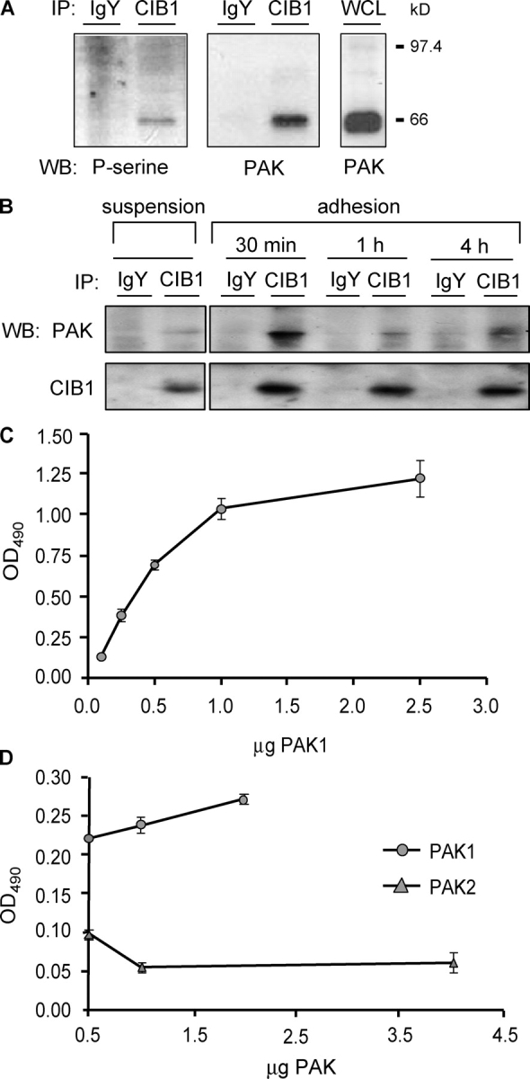
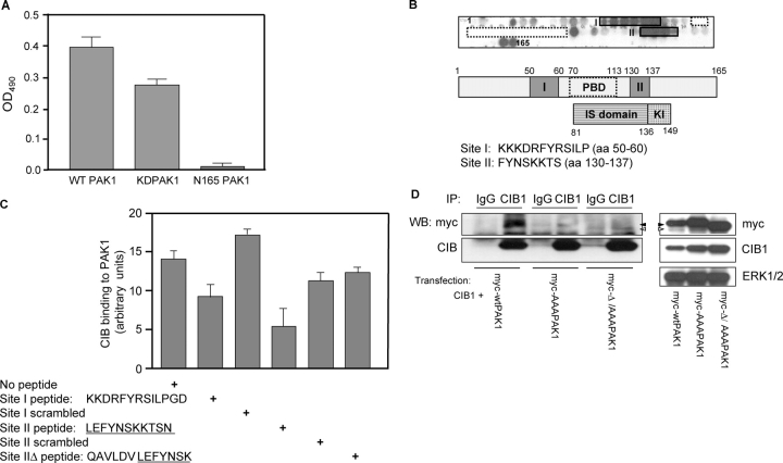
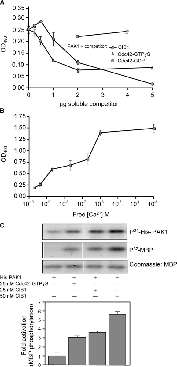
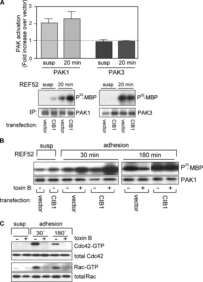
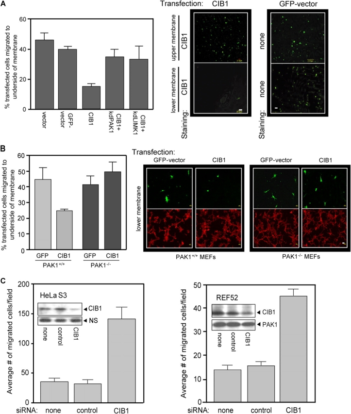
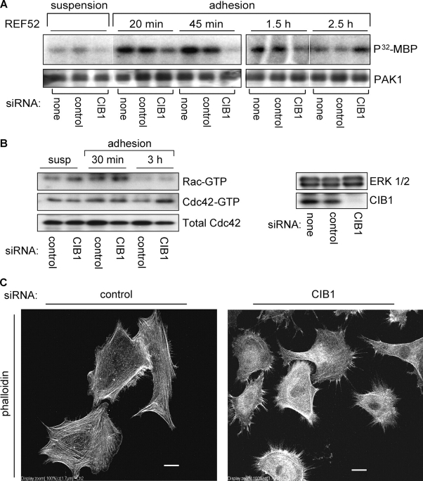
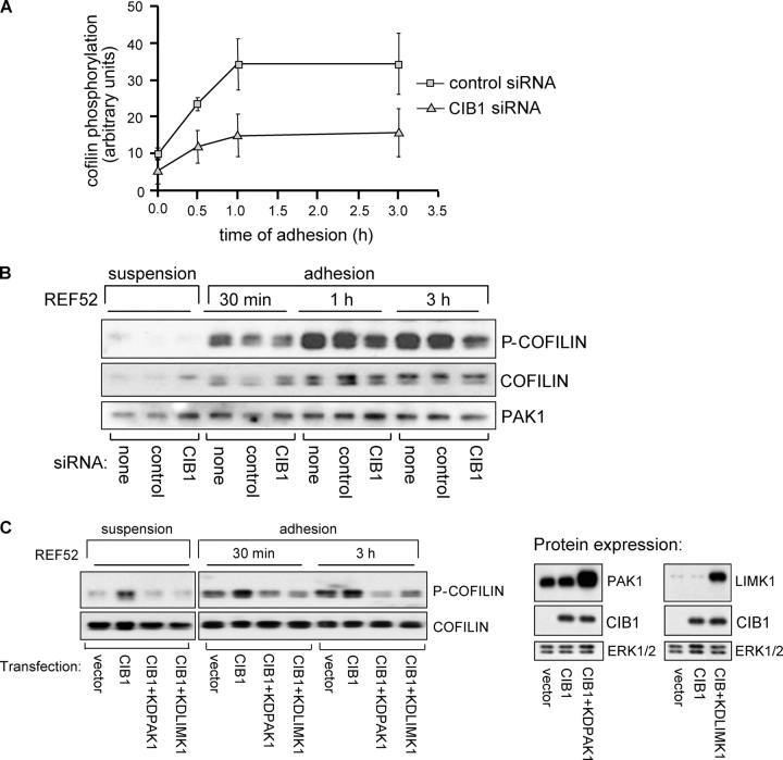
p21-activated kinases (PAKs) regulate many cellular processes, including cytoskeletal rearrangement and cell migration. In this study, we report a direct and specific interaction of PAK1 with a 22-kD Ca2+-binding protein, CIB1, which results in PAK1 activation both in vitro and in vivo. CIB1 binds to PAK1 within discrete regions surrounding the inhibitory switch domain in a calcium-dependent manner, providing a potential mechanism of CIB1-induced PAK1 activation. CIB1 overexpression significantly decreases cell migration on fibronectin as a result of a PAK1-and LIM kinase-dependent increase in cofilin phosphorylation. Conversely, the RNA interference-mediated depletion of CIB1 increases cell migration and reduces normal adhesion-induced PAK1 activation and cofilin phosphorylation. Together, these results demonstrate that endogenous CIB1 is required for regulated adhesion-induced PAK1 activation and preferentially induces a PAK1-dependent pathway that can negatively regulate cell migration. These results point to CIB1 as a key regulator of PAK1 activation and signaling.


Figures

Figure 1.
Figure 1.
CIB1 binds to PAK1 in vivo and in vitro. (A) Immunoprecipitates from platelet lysates were immunoblotted with an antiphosphoserine (left) or anti-PAK (middle) antibody. Whole cell lysates (WCL) were probed with an anti-PAK antibody (right; n ≥ 3 experiments). (B) CIB1 and control IgY immunoprecipitates from lysates of REF52 cells in suspension or adhered to FN were immunoblotted for PAK (top) and CIB1 (bottom). (C) Solid-phase binding assays using immobilized CIB1 and soluble PAK proteins. Increasing concentrations of His-PAK1 were added to wells coated with and without immobilized CIB1. His-PAK1 binding was detected using an anti-His antibody. (D) PAK isozyme–binding specificity was determined by adding GST-PAK1 or GST-PAK2 to immobilized CIB1. PAK binding was detected with an anti-GST antibody. WB, Western blot; IP, immunoprecipitates. Error bars represent SEM.
Figure 2.
Figure 2.
Identification of CIB1-binding sites within the PAK1 NH 2 terminus. (A) CIB1 binds to full-length wild-type GST-PAK1 (wtPAK1) and GST-PAK1 K298A (kdPAK1) but not the NH2-terminal deletion GST-N165-PAK1. Solid-phase binding assays were performed as in Fig. 1 C, and soluble GST-PAK1, GST-PAK1 K298A, and GST-N165-PAK1 were added to wells coated with CIB1. (B) Delineation of the CIB1-binding sites in the PAK1 NH2-terminal sequence by SPOT peptide method. Top and middle panels show two sets of CIB1 reactive spots labeled as sites I and II relative to the PBD- or Rac/Cdc42-binding sites (residues 70–113, dotted boxes). The bottom panel indicates the inhibitory switch (IS) and kinase inhibitor (KI) domains. Sequences of corresponding CIB1-binding sites are below. (C) Inhibition of CIB1 binding to PAK1 with site I and II peptides. Ni-NTA agarose beads loaded with purified His-PAK1 were added to recombinant CIB1 that was preincubated with and without PAK1 peptides corresponding to sites I and II and scrambled sites I and II. Peptide sequences of sites I, II, and IIΔ are shown below. The graph represents densitometry of affinity precipitates probed for CIB1 and normalized to His-PAK1 that was immunoblotted from the same membrane. Data represent SEM from two independent experiments (error bars). (D) Loss of CIB1 binding to site I and II PAK1 mutants. Clarified lysates from HEK293 cells cotransfected with CIB1 and myc wild-type (wt)PAK1, myc-PAK1 siteIAAA (myc-AAAPAK1), or myc-PAK1 siteIΔ/siteIIAAA (myc-Δ/AAA PAK1) were immunoprecipitated with a control IgG or anti-CIB1 antibody and with samples immunoblotted for myc (top) and CIB1 (bottom; n ≥ 4). Western blots of the input expression of CIB1 and myc-tagged wtPAK1 (closed arrowheads), AAAPAK (closed arrowheads), or Δ/AAAPAK1 (open arrowheads).
Figure 3.
Figure 3.
Cdc42 and Ca 2+ affect the CIB1–PAK interaction, and CIB1 stimulates PAK1 activity in vitro. (A) Activated Cdc42-GTPγS or CIB1, but not inactive Cdc42-GDP, competes with immobilized CIB1 for binding to soluble His-PAK1. Soluble His-PAK1 was incubated with increasing concentrations of soluble CIB1 or Cdc42 that was preloaded with GDP or GTPγS before incubation with immobilized CIB1. (B) Determination of Ca2+-dependent binding of His-PAK1 to CIB1. His-PAK1 was diluted in buffer containing 0–5 mM EGTA before addition to immobilized CIB1. Approximate free Ca2+ concentrations were calculated using the MaxChelator program (Bers et al., 1994). (C) Stimulation of recombinant His-PAK1 activity by recombinant CIB1 or Cdc42-GTPγS. His-PAK1 autophosphorylation was assayed in the absence (top) or presence of myelin basic protein (MBP) to detect active kinase (middle). White lines indicate that intervening lanes have been spliced out. Myelin basic protein phosphorylation ([32P]MBP) from densitometry analysis induced by His-PAK1 alone was assigned a value of 1 (bar graph). Data represent means ± SEM (error bars; n = 3).
Figure 4.
Figure 4.
CIB1 specifically stimulates PAK1 activity in vivo independently of small GTPases. (A) CIB1 specifically activates the PAK1 isoform. Endogenous PAK1 (bottom left) and PAK3 (bottom right) were immunoprecipitated from lysates prepared from vector- and CIB1-transfected REF52 cells either held in suspension or replated on FN for 20 min. Immunoprecipitated PAK was subjected to in vitro kinase assays using myelin basic protein (MBP) as substrate. The bar graph (top) depicts [32P]MBP values after normalization for PAK immunoprecipitation. [32P]MBP values from vector control cells were set as 1. Data represent SEM from two independent experiments (error bars). (B) GTPase-independent PAK1 activation. Serum-starved vector and CIB1-transfected REF52 cells held in suspension were treated with or without 100 ng/ml toxin B and either left in suspension or adhered to FN. Cells were lysed at the indicated times, and immunoprecipitated endogenous PAK1 was subjected to in vitro kinase assays using myelin basic protein as substrate. White lines indicate that intervening lanes have been spliced out; n = 3. (C) Efficacy of toxin B treatment was determined by assaying REF52 cell lysates for activated Rac1 and Cdc42 (see PAK1 kinase and Rac/Cdc42 activation assays). Affinity precipitates were analyzed by Western blotting for both Rac1 and Cdc42.
Figure 5.
Figure 5.
CIB1 overexpression inhibits, and endogenous CIB1 depletion increases, cell migration. (A) Serum-starved REF52 cells transfected with GFP vector, control vector, or CIB1 ± kdPAK1 or kdLIMK1 were subjected to haptotactic transwell migration assays toward FN. Transfected cells on either the top membrane (nonmigrating cells) or bottom membrane (migrating cells) were visualized by staining for CIB1 expression (middle). Control migration was visualized by GFP fluorescence (right). Cells overexpressing vector CIB1 ± kdPAK1 or ± kdLIMK1 on the top and bottom membranes were also stained as described in migration assays and were counted. Migration is represented as the percentage of the total number of transfected cells from the upper and lower membranes (left). Data represent means ± SEM (n = 3). (B) MEFs derived from wild-type (PAK+/+) and PAK1-null (PAK−/−) mice were transfected with GFP vector or CIB1. Serum-starved cells were assayed for haptotactic migration toward 3 μg/ml FN, and migration was determined as in A (left). Data represent means ± SEM (n = 4). Right panels show representative images of migrated transfected cells (green, top) and phalloidin staining from the same field (red, bottom). (A and B) Bars, 20 μm. (C) HeLaS3 (left) and REF52 (right) cells were mock transfected or transfected with control or specific CIB1 siRNA and subjected to haptotactic transwell migration assays toward FN (as in A). Data represent means ± SEM (error bars; n = 4 for each cell type). Inset blots show representative endogenous CIB1 protein expression and nonspecific (NS) band or PAK1 expression from the same blot as the loading control.
Figure 6.
Figure 6.
CIB1 is required for adhesion-induced PAK1 activation, and loss of CIB1 disrupts PAK1/GTPase signaling. (A) Mock, control, or specific CIB1 siRNA-transfected REF52 cells were either held in suspension or replated onto FN-coated dishes and were lysed at the indicated times. Immunoprecipitated endogenous PAK1 was subjected to in vitro kinase assays (n = 3). (B) Control or CIB1 siRNA-transfected REF52 cell lysates were assayed for activated and total Rac1 and Cdc42. CIB1 knockdown in REF52 cells was confirmed by immunoblotting cell lysates for endogenous CIB1 expression (right) with ERK as a loading control from the same membrane. (C) REF52 cells transfected with control (left) or CIB1 siRNA (right) were replated on FN for 3 h and were stained with phalloidin. Images are representative of two separate experiments. Bars, 20 μm.
Figure 7.
Figure 7.
CIB1 modulates downstream signaling to cofilin. (A) Lysates were prepared from control or specific CIB1 siRNA-transfected REF52 cells either held in suspension or adhered to FN for the indicated times. Densitometry of p-cofilin levels from lysates that were prepared from control or specific CIB siRNA was normalized to ERK or PAK1 from the same blots. Error bars represent means ± SEM (n = 2). (B) Representative membrane immunoblotted with antibodies against total or phosphorylated cofilin (p-cofilin). The top half of the membrane was also immunoblotted for PAK1 expression (bottom). (C) Lysates prepared from REF52 cells overexpressing empty vector or CIB1 ± kdPAK1 or kdLIMK1 were analyzed for cofilin phosphorylation as in A. The membrane was reprobed for total cofilin (bottom). Lysates from cells expressing control vector, CIB1, or CIB1 coexpressed with kdPAK1 or kdLIMK1 were immunoblotted using anti-CIB1, -PAK1, or -LIMK1 antibodies (middle and right). PAK1 immunoblots show both endogenous PAK1 and overexpressed kdPAK1 (top middle). Immunoblotting for LIMK1 also shows endogenous LIMK1 and overexpressed kdLIMK1 (top right). Middle blots show overexpressed CIB1. Membranes were also probed with an anti-ERK antibody as a loading control (bottom, middle and right). Data represent two separate experiments.

References

    1. Adam, L., R. Vadlamudi, M. Mandal, J. Chernoff, and R. Kumar. 2000. Regulation of microfilament reorganization and invasiveness of breast cancer cells by kinase dead p21-activated kinase-1. J. Biol. Chem. 275:12041–12050. - PubMed
    1. Arber, S., F.A. Barbayannis, H. Hanser, C. Schneider, C.A. Stanyon, O. Bernard, and P. Caroni. 1998. Regulation of actin dynamics through phosphorylation of cofilin by LIM-kinase. Nature. 393:805–809. - PubMed
    1. Bagheri-Yarmand, R., M. Mandal, A.H. Taludker, R.A. Wang, R.K. Vadlamudi, H.J. Kung, and R. Kumar. 2001. Etk/Bmx tyrosine kinase activates Pak1 and regulates tumorigenicity of breast cancer cells. J. Biol. Chem. 276:29403–29409. - PubMed
    1. Bagrodia, S., S.J. Taylor, C.L. Creasy, J. Chernoff, and R.A. Cerione. 1995. Identification of a mouse p21Cdc42/Rac activated kinase. J. Biol. Chem. 270:22731–22737. - PubMed
    1. Bagrodia, S., S.J. Taylor, K.A. Jordon, L. Van Aelst, and R.A. Cerione. 1998. A novel regulator of p21-activated kinases. J. Biol. Chem. 273:23633–23636. - PubMed

Publication types

MeSH terms