. 2005 Aug 1;170(3):487-96.
doi: 10.1083/jcb.200501038.

The epidermal barrier function is dependent on the serine protease CAP1/Prss8

Affiliations

The epidermal barrier function is dependent on the serine protease CAP1/Prss8

Céline Leyvraz et al. J Cell Biol. .

Abstract

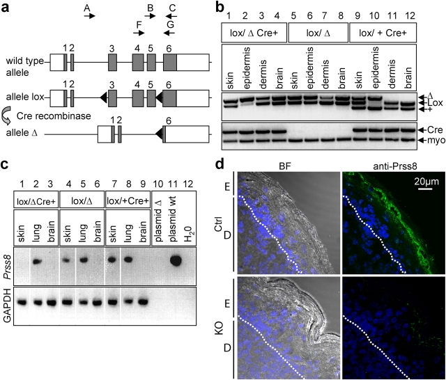
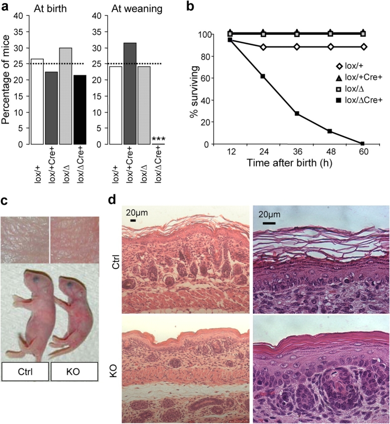
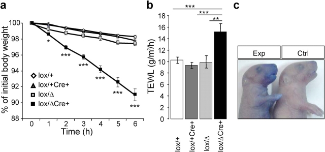
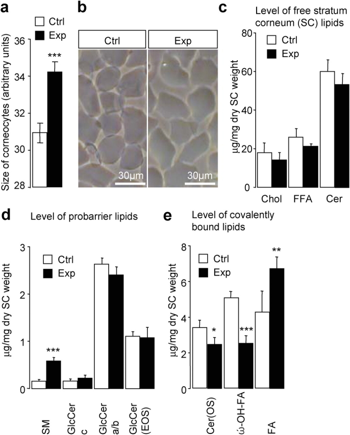
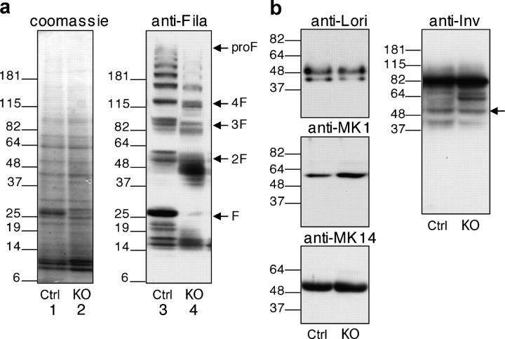
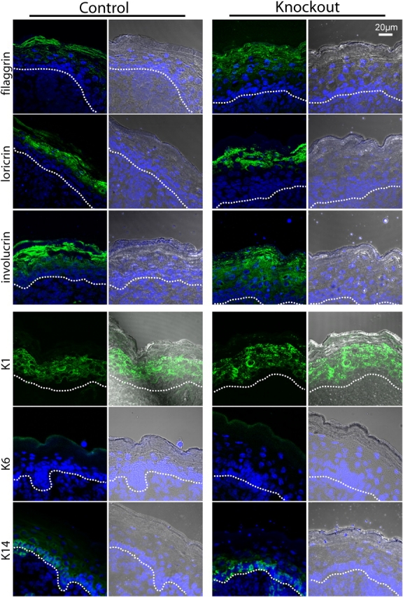
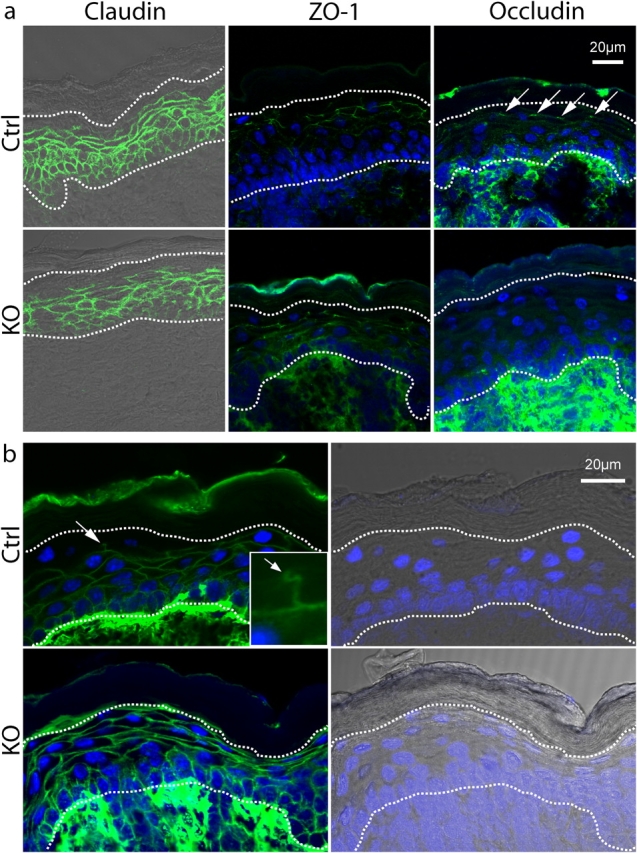
Serine proteases are proteolytic enzymes that are involved in the regulation of various physiological processes. We generated mice lacking the membrane-anchored channel-activating serine protease (CAP) 1 (also termed protease serine S1 family member 8 [Prss8] and prostasin) in skin, and these mice died within 60 h after birth. They presented a lower body weight and exhibited severe malformation of the stratum corneum (SC). This aberrant skin development was accompanied by an impaired skin barrier function, as evidenced by dehydration and skin permeability assay and transepidermal water loss measurements leading to rapid, fatal dehydration. Analysis of differentiation markers revealed no major alterations in CAP1/Prss8-deficient skin even though the epidermal deficiency of CAP1/Prss8 expression disturbs SC lipid composition, corneocyte morphogenesis, and the processing of profilaggrin. The examination of tight junction proteins revealed an absence of occludin, which did not prevent the diffusion of subcutaneously injected tracer (approximately 600 D) toward the skin surface. This study shows that CAP1/Prss8 expression in the epidermis is crucial for the epidermal permeability barrier and is, thereby, indispensable for postnatal survival.


Figures

Figure 1.
Figure 1.
Generation of skin-specific Prss8-deficient mice. (a) Scheme of wild-type, loxP, and Δ allele at the Prss8 gene locus. Coding (gray boxes) and noncoding sequences (white boxes); loxP sites, black triangles. PCR-based genotyping was performed using primers A–C (arrows). For RT-PCR analysis, primers F and G (arrows) were used. (b) PCR analysis with primers A–C distinguishes between wild-type (+; 379 bp), lox (413 bp), and Δ allele (top). Note the shift of the Prss8 lox allele into the Δ allele in epidermis from animals harboring the Cre transgene (lanes 2 and 10). Detection of the Cre transgene using Cre- and myogenin-specific primers (internal control; bottom). (c) RT-PCR analysis using primers F and G demonstrates absence of CAP1/Prss8 mRNA transcripts (309 bp) in the knockout group (lane 1). Wild-type CAP1/Prss8 plasmid (lane 11; positive control). The reaction is controlled by detection of GAPDH transcripts. (d) Immunohistochemistry using the affinity-purified CAP1/Prss8 antibody revealed expression in top layers of the SG and in the transition between SG and SC in the control group (top), but absence of expression in the knockout (KO) group (bottom). BF, bright field; E, epidermis; D, dermis; dotted lines, basal membrane.
Figure 2.
Figure 2.
Early postnatal lethality and skin abnormalities in Prss8lox/ Δ /K14-Cre mice. (a) Distribution of genotypes in the knockout (black bar) and control groups (white, light gray, and dark gray bars) at birth indicated in percentages. From a total of 131 pups (n = 14 litters) that were analyzed shortly after birth, 28 were from the knockout group, 32 were from Prss8 lox/+, 40 were from Prss8 lox/Δ, and 31 were from Prss8 lox/+ /K14-Cre (control group; expected distribution in percentage; dotted line). At weaning, 37 Prss8 lox/+, 37 Prss8 lox/Δ, and 44 Prss8 lox/+ /K14-Cre mice were identified. ***, P < 0.001. (b) Survival curves of knockout (black squares) and control mice (white diamonds and gray squares). Note that no knockout mice survived 60 h after birth. (c) Representative example showing different sizes of knockout (right) and control (left) littermates. (d) Representative skin sections from newborn control (top) and knockout (bottom) littermates stained with hematoxylin and eosin. Staining (left) shows abnormal SC formation and a reduced hair follicle number in knockout skin. Higher magnification (right) reveals an orthokeratotic hyperkeratosis and dysmaturation of hair follicles.
Figure 3.
Figure 3.
Disruption of epidermal barrier function in Prss8lox/ Δ /K14-Cre mice. (a) Dehydration assay over time. Data are presented as percentages of initial body weight in knockout (black squares; n = 5) and control (Prss8 lox/Δ and Prss8 lox/+ /±K14-Cre; white diamonds and gray squares; n = 5–8) mice. *, P < 0.05; ***, P < 0.001. (b) Transepidermal water loss (TEWL) measured on ventral skin is decreased in Prss8 lox/Δ /K14-Cre (black bar; n = 6) pups compared with control pups (Prss8 lox/Δ, light gray bar; n = 9), Prss8 lox/+ /K14-Cre (dark gray bar; n = 11), and Prss8 lox/+ (white bar; n = 15). **, P < 0.01; ***, P < 0.001. Error bars represent SEM. (c) Barrier-dependent dye exclusion assay in knockout (left; n = 8) and control (right; n = 5) pups. Representative photograph revealing dye penetration of all epidermal surfaces, especially ventrally in knockout, but not in control, pups.
Figure 4.
Figure 4.
Enlarged corneocytes and altered SC lipid composition in knockout mice. (a) Mean surface area of cells from control (white bar; n = 15–50 corneocytes; n = 6 mice) and knockout (Exp) mice (black bar; n = 15–50 corneocytes; n = 5 mice). ***, P < 0.001. (b) Morphological appearance of purified cells that are isolated from the control (left) and knockout (right) epidermis. (c) Levels of main free SC lipids. Chol, cholesterol; FFA, free fatty acid; Cer, ceramides. (d) Analysis of probarrier lipids. Note the increased amount of sphingomyelin (SM) in the knockout group (***, P < 0.001), whereas levels of different glucosylceramide fractions (GlcCer c, GlcCer a/b, and GlcCer(EOS); N-acyl fatty acid acylated in ω-OH position) were unaltered. (e) Level of covalently bound lipids. Note the 50% reduced amount of ω-hydroxylated fatty acid (ω-OH–FA; ***, P < 0.001), the decrease in ceramides (Cer[OS]); *, P < 0.05), and the significantly increased fatty acid (FA; **, P < 0.01) levels in knockout (Prss8 lox/Δ /K14-Cre; black bar) versus control (Prss8 lox/+ ; white bar) groups. Each value is the mean of five animals ± SEM (error bars).
Figure 5.
Figure 5.
Aberrant profilaggrin to filaggrin processing in Prss8-deficient epidermis. (a) Equal amounts of protein extracts of control (lanes 1 and 3) and knockout (lanes 2 and 4) epidermis were separated through SDS-PAGE under reducing conditions and either were Coomassie blue stained (loading control; lanes 1 and 2) or subjected to Western blot analysis using the anti-filaggrin antibody (lanes 3 and 4). Note the aberrant profilaggrin to filaggrin processing in the knockout epidermis with a nearly complete loss of 25-kD filaggrin monomers (lane 4). The position of profilaggrin (proF) and its proteolytically-derived products (F; e.g., filaggrin three-domain [3F] and two-domain [2F] intermediates) are indicated. The size of the molecular marker (kD) is also indicated. (b) Western blot analysis on epidermal proteins from control (Ctrl) and knockout (KO) mice after SDS-PAGE separation using differentiation markers (loricrin, 57 kD; K1, 67 kD; K14, 55 kD; and involucrin, 56 kD; indicated by arrow). Equal amounts of the proteins were loaded.
Figure 6.
Figure 6.
Normal distribution of the differentiation markers in CAP1/Prss8-deficient epidermis. Immunofluorescence LSM for filaggrin, loricrin, involucrin, K1, K6, and K14. Nuclei are counterstained with DAPI. Brightfield/DAPI is equally depicted, with the dotted lines indicating the basal membrane. Filaggrin is mainly localized in the SC but starts in the SG. Loricrin staining is observed in the top SG and continues in the SC. Involucrin staining is strong in the SC but is also expressed in the SG and expressed slightly in the stratum spinosum. K1 is localized from the SP to the SG (intermediate epidermis marker). K6 is not detectable in both control and knockout groups. K14, which is a specific marker for the stratum basale, is localized as expected in the two groups.
Figure 7.
Figure 7.
Expression of TJ proteins and TJ permeability assay. (a) Immunofluorescence LSM for claudin-1 (claudin), Zonula occludens 1 (ZO-1), and occludin. For ZO-1 and occludin, the DAPI counterstaining is added. The two dotted lines show the basal membrane (bottom) and the limit SG/SC (top). Claudin staining is localized in the whole epidermis except for the SC and the last layer of SG in both groups. ZO-1 shows a staining in the mid-SG in both groups. In controls, occludin shows a dotted staining, as expected for TJs (arrows), between the second and last layer of the SG. No such staining could be observed in knockouts. (b) TJ permeability assay visualized by LSM. For each genotype, the staining for streptavidin/AlexaFluor488 is shown with DAPI counterstaining, and brightfield/DAPI of the same picture is added for localization. The bottom dotted line indicates the basal membrane, and the top line indicates the limit SG/SC. The top panels show the diffusion of biotin in controls, which is blocked abruptly between the second and last layer of the SG (arrow). The magnification (inset, arrow) indicates the position of TJ. The bottom panel shows the diffusion of biotin in knockout epidermis, which is not blocked by the TJ and, thus, extends up to the SC, where it accumulates.

References

    1. Barel, A.O., and P. Clarys. 1995. Study of the stratum corneum barrier function by transepidermal water loss measurements: comparison between two commercial instruments: Evaporimeter and Tewameter. Skin Pharmacol. 8:186–195. - PubMed
    1. Benavides, F., M.F. Starost, M. Flores, I.B. Gimenez-Conti, J.-L. Guénet, and C.J. Conti. 2002. Impaired hair follicle morphogenesis and cycling with abnormal epidermal differentiation in nackt mice, a cathepsin L-deficient mutation. Am. J. Pathol. 161:693–703. - PMC - PubMed
    1. Brandner, J.M., S. Kief, C. Grund, M. Rendl, P. Houdek, C. Kuhn, E. Tschachler, W.W. Franke, and I. Moll. 2002. Organization and formation of the tight junction system in human epidermis and cultured keratinocytes. Eur. J. Cell Biol. 81:253–263. - PubMed
    1. Brouard, M., M. Casado, S. Djelidi, Y. Barrandon, and N. Farman. 1999. Epithelial sodium channel in human epidermal keratinocytes: expression of its subunits and relation to sodium transport and differentiation. J. Cell Sci. 112:3343–3352. - PubMed
    1. Chen, L.M., X. Zhang, and K.X. Chai. 2004. Regulation of prostasin expression and function in the prostate. Prostate. 59:1–12. - PubMed

Publication types

MeSH terms