. 2005 Aug 1;202(3):445-55.
doi: 10.1084/jem.20051060.

Selective targeting of regulatory T cells with CD28 superagonists allows effective therapy of experimental autoimmune encephalomyelitis

Affiliations

Selective targeting of regulatory T cells with CD28 superagonists allows effective therapy of experimental autoimmune encephalomyelitis

Niklas Beyersdorf et al. J Exp Med. .

Abstract

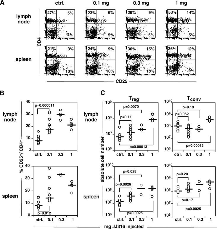
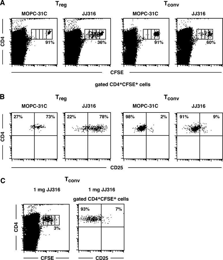
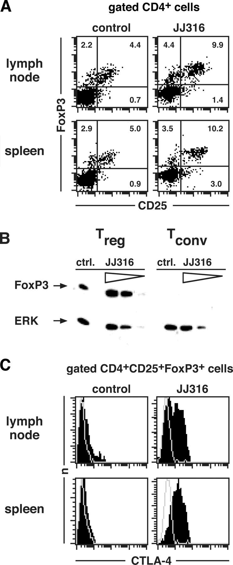
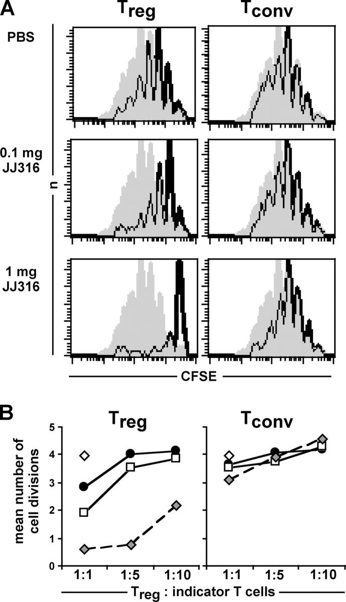
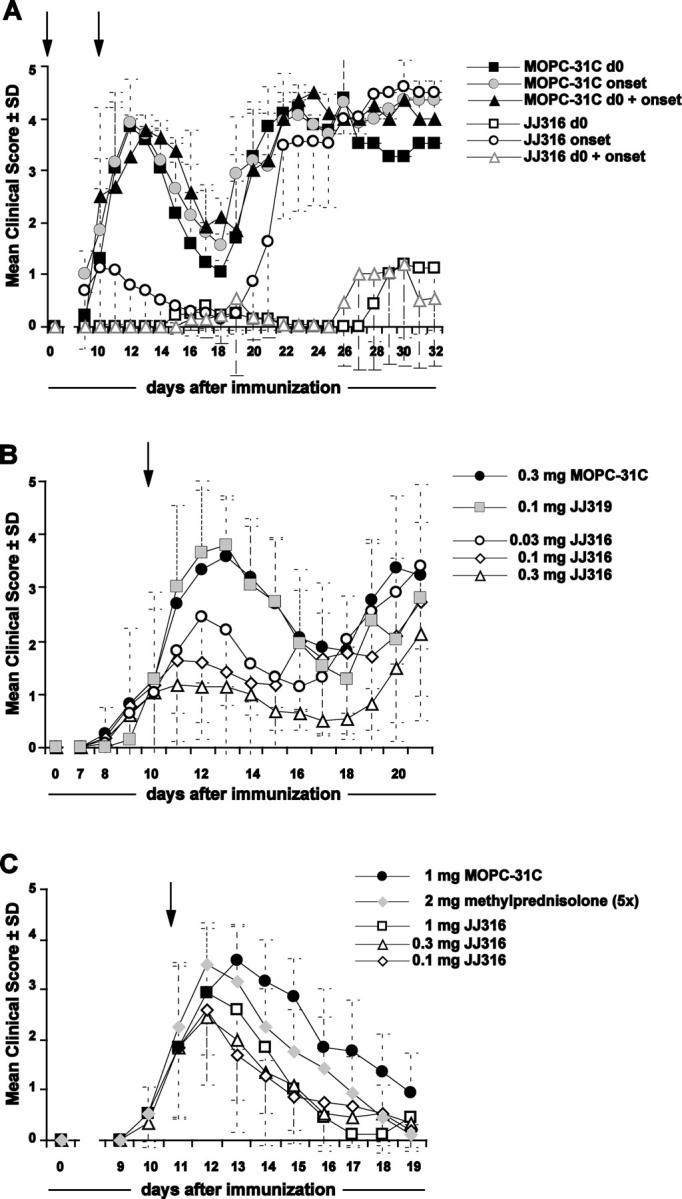
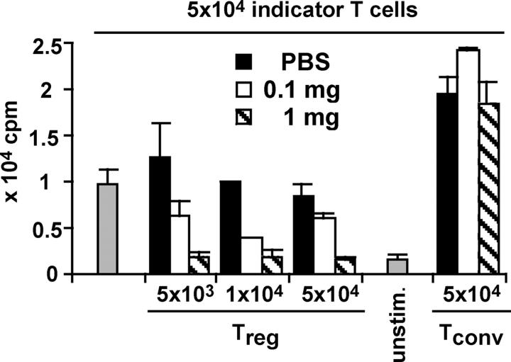
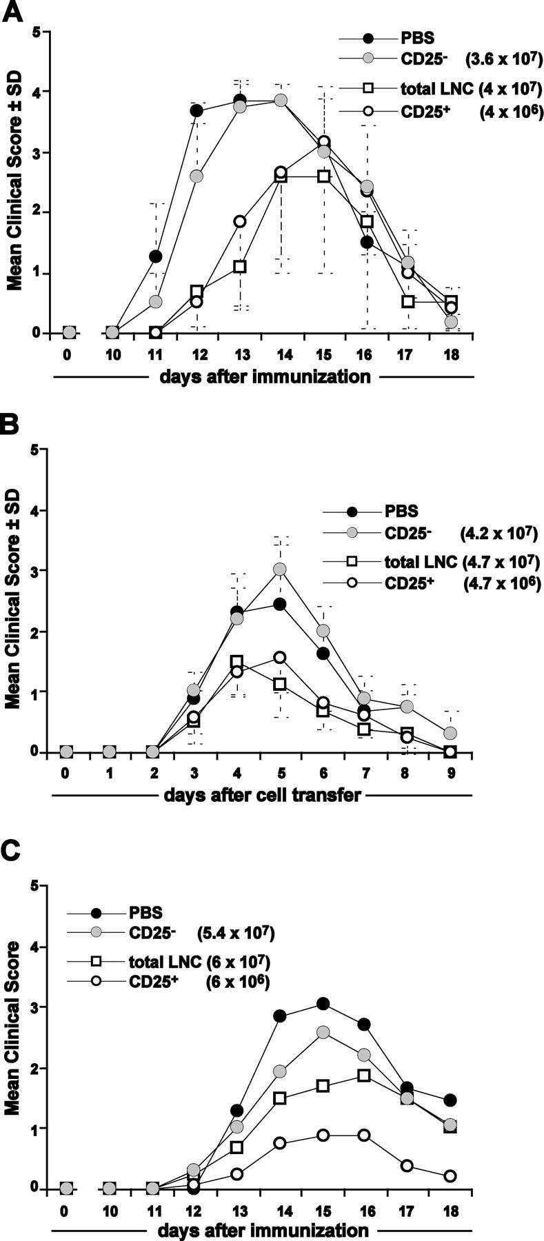
CD4+CD25+ regulatory T cells (T reg cells) play a key role in controlling autoimmunity and inflammation. Therefore, therapeutic agents that are capable of elevating numbers or increasing effector functions of this T cell subset are highly desirable. In a previous report we showed that a superagonistic monoclonal antibody specific for rat CD28 (JJ316) expands and activates T reg cells in vivo and upon short-term in vitro culture. Here we demonstrate that application of very low dosages of the CD28 superagonist into normal Lewis rats is sufficient to induce T reg cell expansion in vivo without the generalized lymphocytosis observed with high dosages of JJ316. Single i.v. administration of a low dose of the CD28 superagonist into Dark Agouti (DA) rats or Lewis rats that suffered from experimental autoimmune encephalomyelitis (EAE) proved to be highly and equally efficacious as high-dose treatment. Finally, we show that T reg cells that were isolated from CD28-treated animals displayed enhanced suppressive activity toward myelin basic protein-specific T cells in vitro, and, upon adoptive transfer, protected recipients from EAE. Our data indicate that this class of CD28-specific monoclonal antibodies targets CD4+CD25+ T reg cells and provides a novel means for the effective treatment of multiple sclerosis and other autoimmune diseases.


Figures

Figure 1.
Figure 1.
In vivo dose-response titration of mAb JJ316. Adult Lewis rats were injected i.v. with the three indicated amounts of mAb JJ316. 3 d after injection, peripheral LNCs and splenocytes were analyzed for the prevalence of CD4+CD25+ and CD4+CD25 cells by FACS analysis. (A) Representative dot plots of CD4 and CD25 expression in lymph nodes and spleens of control or JJ316-treated animals; the figure indicates the percentages of cells in the respective quadrant. (B) The proportion of CD25+ among CD4+ cells is summarized for all animals analyzed. Absolute cell numbers were obtained by multiplying total cell numbers with the relative cell numbers obtained by FACS analysis (C). The plots in (B) and (C) show pooled data obtained on eight different occasions. Each circle represents one animal (i.e., 2 to 12 animals per group). Horizontal bars indicate medians. Mann-Whitney rank sum tests were performed between groups as indicated by the brackets, and the respective P values are given.
Figure 2.
Figure 2.
Direct monitoring of T reg and Tconv cell proliferation upon superagonistic anti-CD28 stimulation in vivo. T reg and Tconv cells were purified from pooled spleens and lymph nodes, labeled with CFSE, and adoptively transferred into syngeneic recipients. 0.1 mg of mAb JJ316 or control mAb MOPC-31C was injected on the following day. CFSE dilution was analyzed 2 d after antibody injection (A). Percentages represent the proportion of undivided cells among transferred cells. The result of one of at least three experiments is shown. (B) CD25 expression on transferred T reg and Tconv cells isolated from MOPC-31C–treated or JJ316-treated animals was determined by counter-staining with anti-CD25 mAb. (C) CFSE dye dilution among, and CD25 expression on, Tconv cells were assessed after stimulation with 1 mg of JJ316.
Figure 3.
Figure 3.
Phenotypic analysis of T reg and Tconv cells from treated animals. 3 d after administration of 1 mg JJ316, lymph node and spleen cells were stained at the cell surface with mAbs against CD4 and CD25, followed by intracellular staining for FoxP3 and CTLA-4. Control animals were injected with PBS only. (A) Representative expression of CD25 and FoxP3 on gated CD4+ cells. The figure indicates the percentages of cells in the respective quadrant. (B) Detection of FoxP3 expression by Western blot analysis. CD4+CD25+ and CD4+CD25 T cells were purified from untreated or JJ316-treated animals (1 mg), and protein lysates from whole cells were generated. FoxP3 expression was detected with a polyclonal rabbit-anti–mouse FoxP3 IgG. Lysates from cells purified after JJ316 treatment also were diluted serially and protein loading was assessed with polyclonal anti–rat ERK-2 Ig. The experiment was repeated with a similar result. (C) Representative anti–CTLA-4 staining profiles of CD4+CD25+FoxP3+ cells are depicted (filled line graphs). Staining specificity was controlled by preincubation with unconjugated anti–CTLA-4 mAb (gray profile). A repeat experiment rendered similar results.
Figure 4.
Figure 4.
Functional analysis of T reg and Tconv cells from JJ316-treated animals ex vivo. (A) Purified T reg and Tconv cells from the lymph nodes of animals treated as indicated 3 d before isolation were cocultured with CFSE-labeled CD4+CD25 T cells from the PBS-treated animal (indicator T cells), and stimulated with costimulatory beads for 5 d. Line graphs show proliferations of indicator T cells in the absence (gray shadow) or the presence of T reg or Tconv cells at a 1:1 ratio (black line). (B) T reg and Tconv cell/indicator T cell ratios of 1:1, 1:5, and 1:10 were determined by translating CFSE dilution profiles into the average number of cell divisions of the indicator T cells after 5 d of culture. The symbols indicate proliferation of indicator T cells in the absence of T reg/Tconv cells (open diamonds) or in the presence of T reg/Tconv cells from the animal treated with PBS (filled circles), 0.1 mg JJ316 (open squares), or 1 mg JJ316 (gray diamonds). The result is representative of two experiments performed.
Figure 5.
Figure 5.
Therapeutic potential of superagonistic CD28-specific mAb JJ316 on active EAE of the Lewis and the DA rat. (A) To induce EAE in DA rats, animals were immunized with 100 μg recombinant human MOG in CFA at day 0. mAb JJ316 (1 mg) was administered i.v. to groups of animals (n = 8) at day 0 (induction of EAE, open squares), day 10 (onset of EAE, open circles), or on days 0 and 10 (open triangles). Control animals (n = 5) were injected with 1 mg of MOPC-31C at the different time points as indicated. Animals were scored for clinical signs of EAE as described in Material and methods. (B) Therapeutic application of 0.3 mg, 0.1 mg, or 0.03 mg of JJ316 after onset of disease (day 10, arrow) protected DA rats (n = 6) immediately from severe EAE. Control animals were injected with 0.3 mg of MOPC-31C or 0.1 mg of the conventional anti-CD28 mAb JJ319. Mann-Whitney U-tests comparing JJ316- and sham-treated rats on days 13 and 14 after disease onset were significant (P < 0.05) for animals receiving 0.03 mg of JJ316 and highly significant (P < 0.01) for animals receiving 0.3 mg of JJ316. (C) Active EAE of the Lewis rat was induced by immunization with gpMBP in CFA in the hind footpad at day 0. On day 11 (arrow), animals received a single injection of 1 mg of an isotype control antibody (filled circles); a single injection of 0.1 mg, 0.3 mg, or 1 mg of mAb JJ316; or were administered 2 mg of methylprednisolone on five consecutive days. P values of Mann-Whitney rank sum tests of clinical scores that compared the isotype-treated group with the JJ316-treated groups on days 13, 14, and 15 were all significant; most were highly significant.
Figure 6.
Figure 6.
mAb JJ316-primed T reg cells suppress the proliferation of gpMBP-specific T cells in vitro. T reg and Tconv cells were isolated from the draining lymph nodes of animals that had been immunized with gpMBP in CFA 9 d before functional analysis. mAb JJ316 (0.1 mg or 1 mg per animal) or PBS was administered 3 d before the in vitro suppression assay. Conventional CD4+CD25 T cells from the PBS-treated animal (gray bars) served as indicator T cells, and were cocultured with T reg or Tconv cells from the animal treated with PBS, 0.1 mg JJ316, or 1 mg of JJ316. T cells were stimulated with gpMBP in the presence of irradiated splenic APCs. A repeat experiment rendered similar results.
Figure 7.
Figure 7.
CD4+CD25+ T reg cells mediate reduced disease severity. Donor animals were immunized with gpMBP in CFA and received 1 mg of mAb JJ316 i.v. 3 d later, draining LNCs were prepared and separated, in part, into CD4+CD25 and CD4+CD25+ cells. Recipient animals received PBS only, 4 (4.7) × 107 total draining LNCs, 4 (4.7) × 106 purified CD4+CD25+ T cells, or 3.6 (4.2) × 107 CD4+CD25 T cells; numbers in parentheses denote the amount of cells transferred in AT-EAE (see Fig. 4 B). EAE was induced in recipient animals by active immunization with gpMBP in CFA (A) or by adoptive transfer of encephalitogenic T cell line cells (B). These experiments were reproduced twice with similar results. Generation of protective CD4+CD25+ T reg cells is related to the application of superagonistic CD28-specific mAb and is antigen priming-independent (C). On the day of immunization with gpMBP in CFA, animals received 6 × 10⁷ JJ316-primed total draining LNCs (open squares) or 6 × 10⁶ separated CD4+CD25+ T cells (open circles). Control treatment consisted of the transfer of 5.4 × 10⁷ CD28 superagonist-activated CD4+CD25 T cells (gray circles) or in the administration of PBS only (black circles).

References

    1. Walker, L.S., and A.K. Abbas. 2002. The enemy within: keeping self-reactive T cells at bay in the periphery. Nat. Rev. Immunol. 2:11–19. - PubMed
    1. Sakaguchi, S., N. Sakaguchi, M. Asano, M. Itoh, and M. Toda. 1995. Immunologic self-tolerance maintained by activated T cells expressing IL-2 receptor alpha-chains (CD25). Breakdown of a single mechanism of self-tolerance causes various autoimmune diseases. J. Immunol. 155:1151–1164. - PubMed
    1. Suri-Payer, E., A.Z. Amar, A.M. Thornton, and E.M. Shevach. 1998. CD4+CD25+ T cells inhibit both the induction and effector function of autoreactive T cells and represent a unique lineage of immunoregulatory cells. J. Immunol. 160:1212–1218. - PubMed
    1. Dieckmann, D., C.H. Bruett, H. Ploettner, M.B. Lutz, and G. Schuler. 2002. Human CD4(+)CD25(+) regulatory, contact-dependent T cells induce interleukin 10-producing, contact-independent type 1-like regulatory T cells. J. Exp. Med. 196:247–253. - PMC - PubMed
    1. Jonuleit, H., E. Schmitt, M. Stassen, A. Tuettenberg, J. Knop, and A.H. Enk. 2001. Identification and functional characterization of human CD4(+)CD25(+) T cells with regulatory properties isolated from peripheral blood. J. Exp. Med. 193:1285–1294. - PMC - PubMed

Publication types

MeSH terms