Comparative Study
. 2005 Aug 9;102(32):11474-9.
doi: 10.1073/pnas.0504388102. Epub 2005 Aug 1.

Cardiac repair with intramyocardial injection of allogeneic mesenchymal stem cells after myocardial infarction

Affiliations
Comparative Study

Cardiac repair with intramyocardial injection of allogeneic mesenchymal stem cells after myocardial infarction

Luciano C Amado et al. Proc Natl Acad Sci U S A. .

Abstract

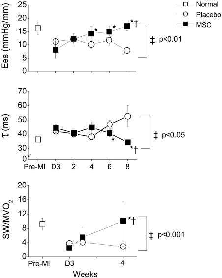
Although clinical trials of autologous whole bone marrow for cardiac repair demonstrate promising results, many practical and mechanistic issues regarding this therapy remain highly controversial. Here, we report the results of a randomized study of bone-marrow-derived mesenchymal stem cells, administered to pigs, which offer several new insights regarding cellular cardiomyoplasty. First, cells were safely injected by using a percutaneous-injection catheter 3 d after myocardial infarction. Second, cellular transplantation resulted in long-term engraftment, profound reduction in scar formation, and near-normalization of cardiac function. Third, transplanted cells were pre-prepared from an allogeneic donor and were not rejected, a major practical advance for widespread application of this therapy. Together, these findings demonstrate that the direct injection of cellular grafts into damaged myocardium is safe and effective in the perii-nfarct period. The direct delivery of cells to necrotic myocardium offers a valuable alternative to intracoronary cell injections, and the use of allogeneic mesenchymal stem cells provides a valuable strategy for cardiac regenerative therapy that avoids the need for preparing autologous cells from the recipient.


Figures

Fig. 1.
Fig. 1.
Engraftment of allogeneic porcine MSCs assessed with MRI. (A) Pseudolong-axis image obtained after MSC injection. Magnetically labeled MSCs appear as hypoenhanced areas (arrows) visible at 2 d and at 1, 4, and 8 weeks after injection. (Right) Bar graph illustrating Feridex retention over 8 weeks. (B) Histologic evaluation of postinfarct myocardium. (Left) Hematoxylin/eosin (H&E) stains obtained from noninjected infarcted area, demonstrating an abundance of inflammatory cells (filled arrows) surrounding the necrotic region (×160 magnification). (Right) H&E and Prussian blue (PB) stains of MSC injection sites, demonstrating the absence of inflammatory cells. Open arrows indicate Feridex (iron)-labeled MSCs.
Fig. 2.
Fig. 2.
Engraftment and differentiation of MSCs. DAPI- and Di-I-labeled MSCs (blue-staining nuclei and red-staining membranes, respectively) and fluorescent muscle-protein-specific antibodies (green). (A) Hematoxylin/eosin (H&E)-stained section and corresponding fluorescent detection of cellular labels. (B) A cluster of MSCs in proximity to host myocardium. Several muscle-specific proteins are detected by immunofluorescence, including α-actinin (C), phospholamban (D), tropomyosin (E), and troponin T (F). Yellow fluorescence indicates colocalization of immunofluorescent antibodies and DiI.
Fig. 3.
Fig. 3.
Impact of MSC injection on myocardial morphology. (A) Representative example of MI scar formation in placebo-treated (Upper) and MSC-treated (Lower) animals at 8 weeks after injury. MSC injection reduces the scar, which is confined to the midmyocardium because of viable tissue in subendocardial and subepicardial zones. (B) Bar graph depicting MI size as the percentage of LV mass in MSC-treated vs. placebo-treated pigs in a randomized study (*, P = 0.008). (C) MSC cardiomyoplasty augments development of new myocardium (Left). At 8 weeks after MI, the subendocardial rim is thicker in the MSC group (arrows). Hematoxylin/eosin (H&E) stain of the subendocardial rim demonstrates cardiomyocytes in both control and MSC (×100 magnification). (D) Short-axis delayed-enhancement images, illustrating the change in infarct size after MSC treatment. At 1 week after MSC injection, there is anteroseptal myocardial necrosis and extensive microvascular obstruction (white arrow in Left). MSC administration leads to myocardial tissue regeneration (red arrows) and reduction of necrotic myocardium (yellow arrows) 8 weeks later (Center). (Left) Corresponding gross pathology. (E) Bar graph, depicting enhanced thickness of subendocardial viable tissue with MSCs (*, P < 0.05 vs. nontreated) after 8 weeks. (F) MI size assessed from MRI delayed hyperenhancement, showing that, in nontreated animals, infarct size does not decrease but is reduced ≈50% with MSC treatment (*, P < 0.05 vs. baseline images obtained 2 d after therapy; †, P < 0.05 vs. nontreated).
Fig. 4.
Fig. 4.
Myocardial regeneration in MSC-treated hearts. Immunohistochemical evaluation of the subendocardial rim of infarcted myocardium 10 d postinjection. (A) Rim of c-Kit-positive myocytes (filled arrow) is visible along the infarct border in both groups and within capillaries (open arrow) in MSC but not in control group. (B) Ki67-positive myocytes present within MSC-treated heart.
Fig. 5.
Fig. 5.
Physiologic impact of MSC therapy after MI. Immediately after MI (D3, day 3), systolic and diastolic cardiac functions are impaired in both groups (P = NS for differences between groups at day 3 after MI). (Top) Ventricular elastance (Ees) declines in placebo-treated pigs but increases dramatically in the MSC group, with complete restoration of Ees to normal levels at 8 weeks (P = NS vs. normal). (Middle) Isovolemic ventricular relaxation (τ) returns to normal in MSC-treated pigs but remains impaired in placebo-treated pigs. (Bottom) The impact of MSC therapy on myocardial mechanoenergetics (SW/MVO2) precedes the restoration of systolic and diastolic cardiac function. During the 4 weeks after MI, SW declines in placebo-treated animals, whereas MVO2 increases, leading to a reduced SW/MVO2 ratio. In MSC-treated pigs, SW increases and MVO2 decreases, resulting in augmented SW/MVO2 and restoration of mechanoenergetic coupling toward normal [P < 0.05 vs. D3, respectively, by repeated measurements ANOVA (†); *, P < 0.05 by Bonferroni; ‡, P < 0.05 between groups by two-way ANOVA].

Similar articles

Cited by

References

    1. Schachinger, V., Assmus, B., Britten, M. B., Honold, J., Lehmann, R., Teupe, C., Abolmaali, N. D., Vogl, T. J., Hofmann, W. K., Martin, H., et al. (2004) J. Am. Coll. Cardiol. 44, 1690-1699. - PubMed
    1. Britten, M. B., Abolmaali, N. D., Assmus, B., Lehmann, R., Honold, J., Schmitt, J., Vogl, T. J., Martin, H., Schachinger, V., Dimmeler, S., et al. (2003) Circulation 108, 2212-2218. - PubMed
    1. Wollert, K. C., Meyer, G. P., Lotz, J., Ringes-Lichtenberg, S., Lippolt, P., Breidenbach, C., Fichtner, S., Korte, T., Hornig, B., Messinger, D., et al. (2004) Lancet 364, 141-148. - PubMed
    1. Assmus, B., Schachinger, V., Teupe, C., Britten, M., Lehmann, R., Dobert, N., Grunwald, F., Aicher, A., Urbich, C., Martin, H., et al. (2002) Circulation 106, 3009-3017. - PubMed
    1. Strauer, B. E., Brehm, M., Zeus, T., Kostering, M., Hernandez, A., Sorg, R. V., Kogler, G. & Wernet, P. (2002) Circulation 106, 1913-1918. - PubMed

Publication types