Comparative Study
. 2005 Aug 9;102(32):11414-9.
doi: 10.1073/pnas.0502240102. Epub 2005 Aug 1.

Drosophila host defense after oral infection by an entomopathogenic Pseudomonas species

Affiliations
Comparative Study

Drosophila host defense after oral infection by an entomopathogenic Pseudomonas species

Nicolas Vodovar et al. Proc Natl Acad Sci U S A. .

Abstract

Drosophila has been shown to be a valuable model for the investigation of host-pathogen interactions. Study of the Drosophila immune response has been hampered, however, by the lack of true Drosophila pathogens. In nearly all studies reported, the bacteria used were directly injected within the body cavity of the insect, bypassing the initial steps of a natural interaction. Here, we report the identification of a previously uncharacterized bacterial species, Pseudomonas entomophila (Pe), which has the capacity to induce the systemic expression of antimicrobial peptide genes in Drosophila after ingestion. In contrast to previously identified bacteria, Pe is highly pathogenic to both Drosophila larvae and adults, and its persistence in larvae leads to a massive destruction of gut cells. Using this strain, we have analyzed the modulation of the larval transcriptome upon bacterial infection. We found that natural infection by Pe induces a dramatic change in larval gene expression. In addition to immunity genes, our study identifies many genes associated with Pe pathogenesis that have been previously unreported.


Figures
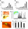
Fig. 1.
Fig. 1.
Expression of antimicrobial peptides after Pe natural infection and persistence within larval gut. (A and B) Natural infection by Pe induces an Imd-dependent systemic immune response. RT-qPCR analysis shows that Pe infection induced sustained Diptericin (A) and Drosomycin (B) expression in wild-type larvae (OrR) but not in relish mutants (monitored only at 0, 3, 6, and 12 h and indicated by an asterisk). 0 h, unchallenged larvae; Dpt, Diptericin; Drs, Drosomycin; rp49, ribosomal protein 49. For each time point, the values represented are the mean and standard deviation of four and three independent experiments for wild-type and relish larvae, respectively. (C) Pe induced local immune response in the gut: histochemical staining of β-galactosidase activity is observed in the anterior midgut at the level of the proventriculus (arrowhead) of wild-type infected larvae that carry the Dpt-lacZ reporter gene. Larvae were collected 24 h after infection. *, endogenous β-galactosidase activity. Similar results were obtained with a Dpt-GFP reporter gene (data not shown). (D) Dpt-lacZ larvae that were sealed at the mouth with a strand of human hair (Inset) were naturally infected by Pe and collected at 12 h. Each bar represents the level of β-galactosidase activity measured in a single larva. Ligatured larvae generally failed to express Dpt-lacZ after exposure to Pe, demonstrating that the digestive tract is the major route of infection for Pe. Unchallenged, untreated larvae; natural infection, larvae infected by Pe; natural infection ligature, ligatured larvae infected by Pe. (E) GFP expressing Pe are observed in the anterior part of the midgut in infected larvae 6 h after infection. A, anterior; P posterior. (F) Bacterial persistence was measured in wild-type (OrR). Bacterial counts were obtained by plating the larval homogenates of five surface-sterilized larvae that were naturally infected with a rifampicin-resistant strain of Pe and its gacA::Tn5 derivative on LB medium containing rifampicin (100 μg/ml). The number of colony-forming units (cfu) per larva obtained at each time point after infection represents the mean of three independent measurements.
Fig. 2.
Fig. 2.
Natural infection by Pe kills both Drosophila larvae and adults. (A) Wild-type (OrR) and relish larvae were naturally infected by Pe. Pe infections kill 70% of wild-type larvae within 48 h. relish larvae were more susceptible to Pe infection (90% lethality at 48 h) compared with OrR larvae. (B) Wild-type (OrR) and relish adult flies were naturally infected by Pe. Pe infection kills 70% of wild-type adults within 4 days. relish flies were more susceptible than wild-type to Pe infection. Unchallenged relish larva and flies survived as wild type (data not shown). (C) Pe ingestion induces food uptake blockage. The medium of unchallenged larvae is kneaded (Left, open arrowhead) contrary to what observed in infected larvae (Right, filled arrowhead). (D) Infection by Pe often induces melanization at the level of the surface of the proventriculus visible in living larvae or on dissected gut (Inset).
Fig. 3.
Fig. 3.
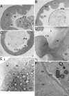
Pe infection provokes a strong perturbation of the Drosophila larval midgut. Transversal sections of larval anterior midgut collected at 6 h (AD)or 12 h (E and F) after natural infection by Pe (BD and F) or a gacA::Tn5 Pe avirulent derivative (A and E) were analyzed. (AC) Semithin sections were observed under bright field. (DF) Ultra-thin sections were observed by transmission electron microscopy. At 6 h after Pe infection, the mucus that protects the digestive epithelium was absent (compare B with A), and the gut cells showed extrusion of cell materials into the lumen (C and D). At 12 h after infection, the cells display abnormal microvilli (compare F with E). M, mucus; L, lumen; m, microvilli; PM, peritrophic matrix; Pe, P. entomophila; CE, extrusion of cell materials; am, abnormal microvilli; EC, epithelial cell. (Scale bar: DF, 1 μm; AC, 5 μm.)
Fig. 4.
Fig. 4.
General statistics on the larval genes regulated after natural infection and septic injury. The graph shows the number of genes induced or repressed after natural infection by Pe (white), Ecc15 (black) and after septic injury with a mixture of E. coli and M. luteus (SI, gray). The 57 genes modulated by Pe, Ecc15, and septic injury belong to the core of immune responsive gene. The 92 genes modulated by both Ecc15 and Pe but not septic injury constitute the core of natural infection specific genes. The 27 genes modulated only by the septic injury may play a role in wound healing and response to Gram-positive bacterial infection.
Fig. 5.
Fig. 5.
Examples of genes regulated by the different types of immune challenge. The fold change after septic injury and natural infection compared with uninfected larvae for selected genes is shown. Time intervals after infection are indicated in hours on the top. The GO IDs are references to entries in the geneontology index of molecular functions and biological processes (www.geneontology.org). For simplicity, the values for the gacA::Tn5 infection have been removed. The fold change color code is indicated at the bottom right.

References

    1. Tzou, P., De Gregorio, E. & Lemaitre, B. (2002) Curr. Oinp. Microbiol. 5, 102-110. - PubMed
    1. Hultmark, D. (2003) Curr. Opin. Immunol. 15, 12-19. - PubMed
    1. De Gregorio, E., Spellman, P. T., Rubin, G. M. & Lemaitre, B. (2001) Proc. Natl. Acad. Sci. USA 98, 12590-12595. - PMC - PubMed
    1. Irving, P., Troxler, L., Heuer, T. S., Belvin, M., Kopczynski, C., Reichhart, J.-M., Hoffmann, J. A. & Hetru, C. (2001) Proc. Natl. Acad. Sci. USA 98, 15119-15124. - PMC - PubMed
    1. Boutros, M., Agaisse, H. & Perrimon, N. (2002) Dev. Cell 3, 711-722. - PubMed

Publication types

MeSH terms

Substances

Associated data