Comparative Study
. 2005 Aug 9;102(32):11438-43.
doi: 10.1073/pnas.0501420102. Epub 2005 Aug 1.

B7-DC cross-linking restores antigen uptake and augments antigen-presenting cell function by matured dendritic cells

Affiliations
Comparative Study

B7-DC cross-linking restores antigen uptake and augments antigen-presenting cell function by matured dendritic cells

Suresh Radhakrishnan et al. Proc Natl Acad Sci U S A. .

Retraction in

Abstract

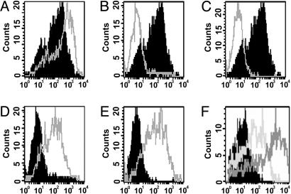
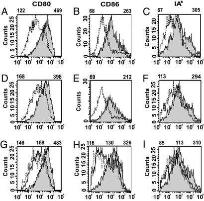
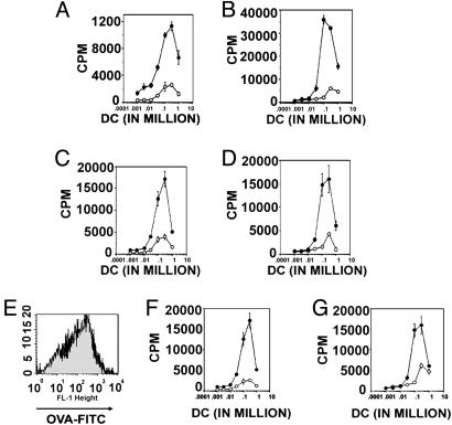
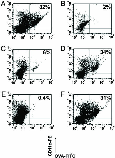
Dendritic cells (DCs) are classified in two states: immature DCs (iDCs), which perform sentinel functions, sampling for antigen and danger signals, and mature DCs (mDCs), which exhibit enhanced antigen-presenting functions but are no longer capable of acquiring antigen. We now describe DCs with a different activation phenotype: cells having the strong antigen-presenting functions of mDCs and the antigen-acquiring functions of iDCs. We have described an antibody that binds the costimulatory molecule B7-DC and activates DCs. The resulting phenotype is distinct from iDCs or mDCs matured by using Toll-like receptor (TLR) agonists. Ability to take up antigen increases, while expression of B71/2 costimulatory and MHC molecules remains unchanged. DCs matured with TLR agonists and then superactivated through B7-DC exhibit a previously unrecognized phenotype. These DCs recover the ability to take up antigen, which is normally lost after treatment with TLR-3 and TLR-9 agonists, yet retain the high expression of costimulatory and MHC molecules and strong antigen-presenting functions of mDCs. Immunization using TLR agonists and B7-DC XAb (cross-linking antibody) together as adjuvant resulted in substantially increased cytolytic T cell responses, particularly when minimal peptide antigens were used. By stimulating DCs with two distinct activation signals, a previously unrecognized phenotype exhibiting augmented antigen-presenting functions was obtained.


Figures

Fig. 1.
Fig. 1.
B7-DC XAb induces uptake of antigen by CD11c+ cells in vitro. DCs were prepared from bone marrow-derived precursors after culture with granulocyte–macrophage colony-stimulating factor and IL-4. Day-5 cultures of DCs were either not treated or pulsed with the TLR agonists CpG-ODN or poly I:C. On day 6, cells received either sHIgM12 B7-DC XAb or isotype control antibody sHIgM39 and were pulsed with FITC-labeled OVA. Accumulation of FITC in washed cells was assessed by flow cytometry on day 7. Histograms represent FITC incorporation by CD11c+ cells. (A) No treatment on day 5, isotype control antibody (black) or B7-DC XAb (gray). (B) No treatment on day 5, isotype control antibody on day 6 (black); CpG-ODN on day 5 and isotype control antibody on day 6 (gray). (C) No treatment on day 5 and isotype control antibody on day 6 (black); poly I:C on day 5 and isotype control antibody on day 6 (gray). (D) CpG-ODN on day 5 and isotype control antibody (black) or B7-DC XAb (gray) on day 6. (E) poly I:C on day 5 and isotype control antibody (black) or B7-DC XAb (gray) on day 6. (F) CpG-ODN on day 5; IgG isotype control antibody (black), anti-CD40 IgG antibody (light gray), or B7-DC XAb (dark gray) on day 6.
Fig. 2.
Fig. 2.
B7-DC cross-linking does not induce CD80, CD86, and class II expression by DCs. DCs were evaluated for expression of the costimulatory molecule CD80, CD86, and IAb expression. Mean fluorescent intensity (MFI) of histograms of cells gated for CD11c expression is displayed above each panel. (A) Day-7 CD80 expression on DCs not treated (dotted line) or treated with CpG-ODN (gray) on day 5, followed by treatment with IgM isotype control antibody on day 6. (B) CD86 expression on cells treated as in A. (C) IAb expression on cells treated as in A.(D, E, and F) CD80, CD86, and IAb expression as in A, B, and C, respectively, except that DCs were treated with sHIgM12 B7-DC XAb on day 6 instead of isotype control IgM antibody. (G, H, and I) CD80, CD86, and IAb expression, respectively, of DCs treated on day 6 only with IgG isotype control antibody (solid line), B7-DC XAb (dotted line), or IgG anti-CD40 antibody (gray). Left MFI value is for isotype control, center MFI value is for B7-DC XAb, and right MFI value is for anti-CD40.
Fig. 3.
Fig. 3.
Treatment of mDCs with B7-DC XAb at the time of antigen pulsing restores antigen-presenting ability. Day-7 DCs from treatment groups shown in Fig. 1 were cocultured with naïve T cells from OT-I and OT-II transgenic mice to evaluate antigen-presenting function. Incorporation of [3H]thymidine was used to evaluate T cell activation. 3H incorporation was evaluated on day 10 (day 3 of coculture with T cells). (A) DCs that received isotype control antibody (open circles) or B7-DC XAb (filled circles) along with soluble OVA on day 6 of culture coincubated for 3 days with OT-I T cells. (B) DCs that received isotype control antibody (open circles) or B7-DC XAb (filled circles) along with soluble OVA on day 6 of culture coincubated for 3 days with OT-II T cells. (C) DCs treated with poly I:C on day 5, isotype control antibody (open circles) or B7-DC XAb (filled circles) and OVA on day 6, and then cocultured with OT-I T cells on day 7. (D) DCs treated with poly I:C on day 5, isotype control antibody (open circles) or B7-DC XAb (filled circles) and OVA on day 6, and then cocultured with OT-II T cells on day 7. (E) Comparison of FITC-OVA incorporation in 7-day DCs that received no treatment on day 5 and isotype control antibody plus OVA on day 6 (black) with DCs that received poly I:C treatment on day 5 and B7-DC XAb and OVA on day 6 (gray). (F) T cell stimulation by DCs from E cocultured with OT-I cells. (G) T cell stimulation by DCs from E cocultured with OT-II cells.
Fig. 4.
Fig. 4.
B7-DC cross-linking induces antigen uptake in lymph node DCs. Animals received i.v. treatments with 10 μg of isotype control antibody (sHIgM39), B7-DC XAb (sHIgM12), or CD40-binding antibody (1C10) 1 day before and on the day of intradermal challenge with 100 μg of FITC-OVA. The draining lymph nodes and contralateral lymph nodes were excised and assessed for FITC incorporation by CD11c+ cells. (A) Draining lymph node cells from animals treated systemically with B7-DC XAb. (B) Contralateral lymph node cells from animals treated systemically with B7-DC XAb. (C) Draining lymph node cells from animals treated systemically with isotype control antibody. (D) Draining lymph node cells from animals treated systemically with CD40-binding antibody. (E) Draining lymph node cells from animals treated systemically with B7-DC XAb and locally at the time of intradermal challenge with FITC-OVA with 10 μg of a rat anti-B7-DC IgG2a antibody. (F) Draining lymph node cells from animals treated systemically with CD40-binding antibodies and locally at the time of intradermal challenge with FITC-OVA with 10 μg of a rat anti-B7-DC IgG2a antibody.
Fig. 5.
Fig. 5.
Stimulation through TLR together with B7-DC cross-linking induces enhanced responses to protein and peptide antigens. Animals received either whole OVA (A and B) or a mixture of the peptides SIINFEKL and PADRE (C and D) in IFA s.c. The animals received no further treatment, a series of four daily treatments with CpG-ODN beginning on the day of antigen challenge, a series of three daily i.v. B7-DC XAb or isotype control antibody challenges beginning 1 day before antigen challenge, or a combination of the treatments. CTLs were harvested from the draining lymph nodes 7 days after antigen challenge and evaluated directly in a 51Cr-release assay. Treatment groups shown are as follows: open circles, CTLs from animals treated with isotype control antibody; filled circles, CTLs from animals treated with B7-DC XAb; squares, CTLs from animals treated with CpG-ODN; triangles, CTLs from animals receiving a combination of B7-DC XAb and CpG-ODN treatments. (A) CTL assay using OVA-immunized T cells and OVA-expressing EG7 cells as targets. (B) CTL assay using T cells in A against EL4 cells that do not express OVA antigens. (C) CTL assay using T cells immunized with a SIINFEKL–PADRE mixture and EG7 targets. (D) CTL assay using T cells described in C against EL4 targets.

References

    1. Banchereau, J., Briere, F., Caux, C., Davoust, J., Lebecque, S., Liu, Y.-J., Pulendran, B. & Palucka, K. (2000) Annu. Rev. Immunol. 18, 767-811. - PubMed
    1. Heath, W. R., Belz, G. T., Behrens, G. M., Smith, C. M., Forehan, S. P., Parish, I. A., Davey, G. M., Wilson, N. S., Carbone, F. R. & Villadangos, J. A. (2004) Immunol. Rev. 199, 9-26. - PubMed
    1. Guermonprez, P., Valladeau, J., Zitvogel, L., Thery, C. & Amigorena, S. (2002) Annu. Rev. Immunol. 20, 621-667. - PubMed
    1. Bhardwaj, N., Bender, A., Gonzalez, N., Bui, L. K., Garrett, M. C. & Steinman, R. M. (1995) Adv. Exp. Med. Biol. 378, 375-379. - PubMed
    1. Latchman, Y., Wood, C. R., Chernova, T., Chaudhary, D., Borde, M., Chernova, I., Iwai, Y., Long, A. J., Brown, J. A., Nunes, R. et al. (2001) Nat. Immunol. 2, 261-268. - PubMed

Publication types

Substances