Review
. 2005 Aug;115(8):1972-83.
doi: 10.1172/JCI26031.

Thyrotropin receptor-associated diseases: from adenomata to Graves disease

Affiliations
Review

Thyrotropin receptor-associated diseases: from adenomata to Graves disease

Terry F Davies et al. J Clin Invest. 2005 Aug.

Abstract

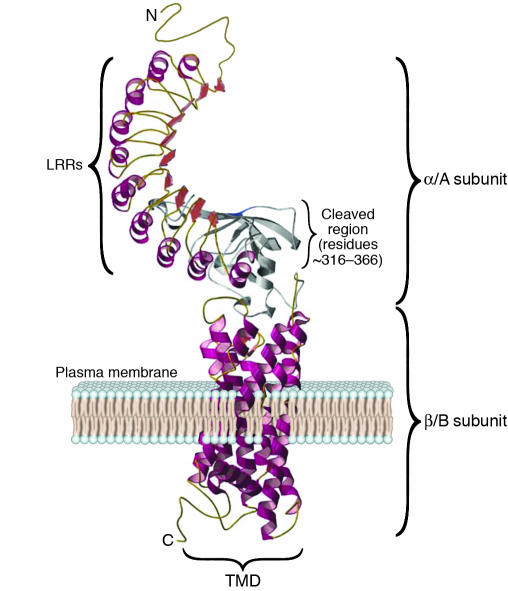
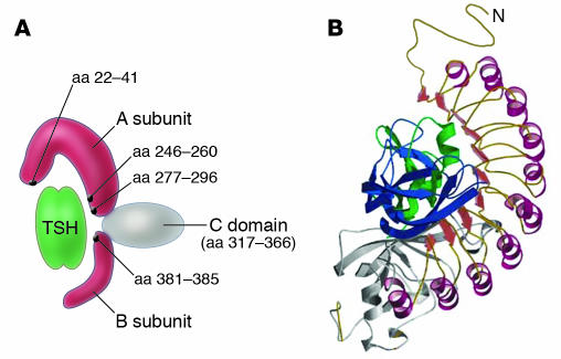
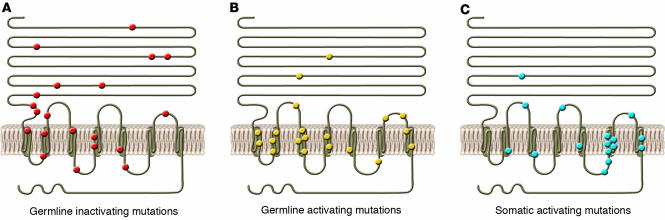
The thyroid-stimulating hormone receptor (TSHR) is a G protein-linked, 7-transmembrane domain (7-TMD) receptor that undergoes complex posttranslational processing unique to this glycoprotein receptor family. Due to its complex structure, TSHR appears to have unstable molecular integrity and a propensity toward over- or underactivity on the basis of point genetic mutations or antibody-induced structural changes. Hence, both germline and somatic mutations, commonly located in the transmembrane regions, may induce constitutive activation of the receptor, resulting in congenital hyperthyroidism or the development of actively secreting thyroid nodules. Similarly, mutations leading to structural alterations may induce constitutive inactivation and congenital hypothyroidism. The TSHR is also a primary antigen in autoimmune thyroid disease, and some TSHR antibodies may activate the receptor, while others inhibit its activation or have no influence on signal transduction at all, depending on how they influence the integrity of the structure. Clinical assays for such antibodies have improved significantly and are a useful addition to the investigative armamentarium. Furthermore, the relative instability of the receptor can result in shedding of the TSHR ectodomain, providing a source of antigen and activating the autoimmune response. However, it may also provide decoys for TSHR antibodies, thus influencing their biological action and clinical effects. This review discusses the role of the TSHR in the physiological and pathological stimulation of the thyroid.


Figures

Figure 1
Figure 1
TSHR structure. This computer model of the TSHR shows the 7 TMDs (spirals) embedded within the plasma membrane and a short cytoplasmic tail, which together make up the β/B subunit. The unique 50-aa–long cleaved region (about 316–366 aa) is shown in gray. Forming a long array, the 9 LRRs, each consisting of 20–24 aa, are depicted as spirals (α helices and β pleated sheets) on the ectodomain of the receptor and make up the major portion of the α/A subunit. The LRRs have a characteristic horseshoe shape with a concave inner surface. C, C-terminus; N, N-terminus. Figure adapted with permission from Thyroid (28).
Figure 2
Figure 2
The TSH-binding pocket and TSHR antibody epitopes. (A) Schematic representation of the TSHR ectodomain showing the major regions (black dots) of TSH binding. Figure adapted with permission from Thyroid (24). (B) Model of the TSH-binding pocket, with TSH ligand making contact with the epitopes within and outside of the LRRs. Figure adapted with permission from Thyroid (28).
Figure 3
Figure 3
Schematic representation of the stages of development of the thyroid follicular cells and the expression of relevant genes. At mouse E8 (E20 in human), the median thyroid bud appears as a thickening in the floor of the pharynx and expresses a combination of transcription factors such as PAX8, the transcription factor essential for the thyrocyte promoter activation of TPO; Tg and NIS; and TTF1 and TTF2, responsible for morphogenesis of the thyroid gland and maintenance of the thyrocyte cell type. At about E13.5 in mouse and E50 in human, the thyroid diverticulum starts its migration from the pharyngeal floor and reaches its definitive pretracheal position. By E14 (E60 in human), the thyroid follicular cell (TFC) precursors express the TSHR. By E15.5, the thyroid follicular organization appears with the expression of a series of proteins that are essential for thyroid hormone biosynthesis, including TPO, Tg, and NIS. Figure modified with permission from Clinical Genetics (47).
Figure 4
Figure 4
Examples of mutation in human and mouse TSHR. The locations of constitutively germline inactivating (A), germline activating (B), and somatic activating (C) mutations are represented. Most activating mutations (shown in B and C) have been localized to exon 10, which codes for the transmembrane and cytoplasmic regions of the receptor. Figure adapted with permission from The New England Journal of Medicine (S62).
Figure 5
Figure 5
Gestational thyrotoxicosis. Shown here is the inverse relationship between serum hCG and TSH levels in early pregnancy. The level of TSH falls as thyroid function increases. Simultaneously, hCG levels increase. Adapted with permission from The Journal of Clinical Endocrinology and Metabolism (87).
Figure 6
Figure 6
A hypothetical model simplified to explain the effect of structural changes in the TSH-binding site by diverse TSHR antibodies. The TSH-binding pocket, represented by the LRRs, is shown by spirals representing the α helix and the β pleated sheets represented by the wide red arrows. The gray region represents the unique cleaved region (316–366 aa) of the receptor. (A) Epitope A1 represents the site where thyroid-stimulating antibodies bind in part to the LRRs, bringing about a structural change in the receptor that leads to signal transduction; Epitope A2 represents a similar competing site, where TSH-blocking antibodies bind (both illustrated as a best fit). (B) Epitope B is the least common site, where TSHR-blocking antibodies may bind but do not compete with antibodies binding to Epitope A. They bind in part to the LRR region but do not bring about the required structural change for signal transduction yet are still able to hinder TSH binding to this site (illustrated as a good fit). (C) Epitope C is where neutral antibodies bind to the cleaved region and/or the N terminus of the TSHR ectodomain, bringing no appropriate structural alteration to the TSHR and thus leaving the LRR region free for TSH, and other TSHR antibodies, to bind. Thus, neutral antibodies result in no signal transduction and do not block TSH binding (illustrated as no fit).

References

    1. Davies TF, Marians R, Latif R. The TSH receptor reveals itself. J. Clin. Invest. 2002;110:161–164. doi:10.1172/JCI200216234. - PMC - PubMed
    1. Duprez L, et al. Pathology of the TSH receptor. J. Pediatr. Endocrinol. Metab. 1999;12(Suppl. 1):295–302. - PubMed
    1. Rapoport B, Chazenbalk GD, Jaume JC, McLachlan SM. The thyrotropin (TSH) receptor: interaction with TSH and autoantibodies [review] Endocr. Rev. 1998;19:673–716. - PubMed
    1. Kajita Y, Rickards CR, Buckland PR, Howell RD, Rees Smith B. Analysis of thyrotropin receptors by photoaffinity labeling. Orientation of receptor subunits in the cell membrane. Biochem. J. 1985;227:413–420. - PMC - PubMed
    1. Parmentier M, et al. Molecular cloning of the thyrotropin receptor. Science. 1989;246:1620–1622. - PubMed

Publication types

Substances