Review
. 2005 Aug;115(8):1990-9.
doi: 10.1172/JCI25505.

Inherited disorders of voltage-gated sodium channels

Affiliations
Review

Inherited disorders of voltage-gated sodium channels

Alfred L George Jr. J Clin Invest. 2005 Aug.

Abstract

A variety of inherited human disorders affecting skeletal muscle contraction, heart rhythm, and nervous system function have been traced to mutations in genes encoding voltage-gated sodium channels. Clinical severity among these conditions ranges from mild or even latent disease to life-threatening or incapacitating conditions. The sodium channelopathies were among the first recognized ion channel diseases and continue to attract widespread clinical and scientific interest. An expanding knowledge base has substantially advanced our understanding of structure-function and genotype-phenotype relationships for voltage-gated sodium channels and provided new insights into the pathophysiological basis for common diseases such as cardiac arrhythmias and epilepsy.


Figures

Figure 1
Figure 1
Structure and genomic location of human NaVChs. (A) Simple model representing transmembrane topology of α and β NaVCh subunits. Structural domains mediating key functional properties are labeled. (B) Chromosomal location of human genes encoding α (red) and β (blue) subunits across the genome. An asterisk next to the gene name indicates association with an inherited human disease. A double asterisk indicates association with murine phenotypes.
Figure 2
Figure 2
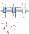
Functional properties of NaVChs. (A) Schematic representation of an NaVCh undergoing the major gating transitions. (B) Voltage-clamp recording of NaVCh activity in response to membrane depolarization. Downward deflection of the current trace (red) corresponds to inward movement of Na+.
Figure 3
Figure 3
A common form of defective inactivation exhibited by mutant NaVChs associated with hyperkalemic periodic paralysis, long QT syndrome, and inherited epilepsy. The defect is caused by incomplete closure of the inactivation gate (left panel) resulting in an increased level of persistent current (right panel, red trace) as compared with NaVChs with normal inactivation (black trace).
Figure 4
Figure 4
Differences between normal and myotonic muscle action potentials. (A) Generation of action potential spikes during electrical stimulation (horizontal blue line and square wave) of a normal muscle fiber. Contraction occurs during action potential firing, followed by muscle relaxation when stimulation ceases. (B) Action potentials in myotonic muscle during and immediately after electrical stimulation. An afterdepolarization triggers spontaneous action potentials that fire after termination of the electrical stimulus (myotonic activity).
Figure 5
Figure 5
Electrophysiological basis for LQTS. (A) Relationship of surface ECG (top) with a representative cardiac action potential (bottom). The QT interval approximates the action potential duration. Individual ionic currents responsible for different phases of the action potential are labeled. (B) Prolongation of the QT interval and corresponding abnormal cardiac action potential (blue) resulting from persistent sodium current. ICa, calcium current; IK1, inward rectifier current; IKr, rapid component of delayed rectifier current; IKs, slow component of delayed rectifier current; INa, sodium current; ITO, transient outward current.
Figure 6
Figure 6
Electrophysiological basis for Brugada syndrome. (A) Comparison of endocardial and epicardial action potentials in normal heart. The epicardial action potential is shorter because of large transient outward current. (B) Endocardial and epicardial action potentials in Brugada syndrome. Reduced sodium current causes disproportionate shortening of epicardial action potentials with resulting exaggeration of the transmural voltage gradient (horizontal double arrow).

References

    1. Catterall WA. Cellular and molecular biology of voltage-gated sodium channels [review] Physiol. Rev. 1992;72(Suppl. 4):S15–S48. - PubMed
    1. Catterall WA. From ionic currents to molecular mechanisms: the structure and function of voltage-gated sodium channels. Neuron. 2000;26:13–25. - PubMed
    1. Whitaker WR, et al. Distribution of voltage-gated sodium channel alpha-subunit and beta-subunit mRNAs in human hippocampal formation, cortex, and cerebellum. J. Comp. Neurol. 2000;422:123–139. - PubMed
    1. Trimmer JS, et al. Primary structure and functional expression of a mammalian skeletal muscle sodium channel. Neuron. 1989;3:33–49. - PubMed
    1. George AL, Komisarof J, Kallen RG, Barchi RL. Primary structure of the adult human skeletal muscle voltage-dependent sodium channel. Ann. Neurol. 1992;31:131–137. - PubMed

Publication types