

A mouse model of juvenile hemochromatosis
- PMID: 16075059
- PMCID: PMC1180543
- DOI: 10.1172/JCI25049
A mouse model of juvenile hemochromatosis
Abstract
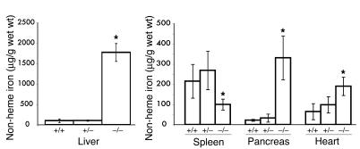
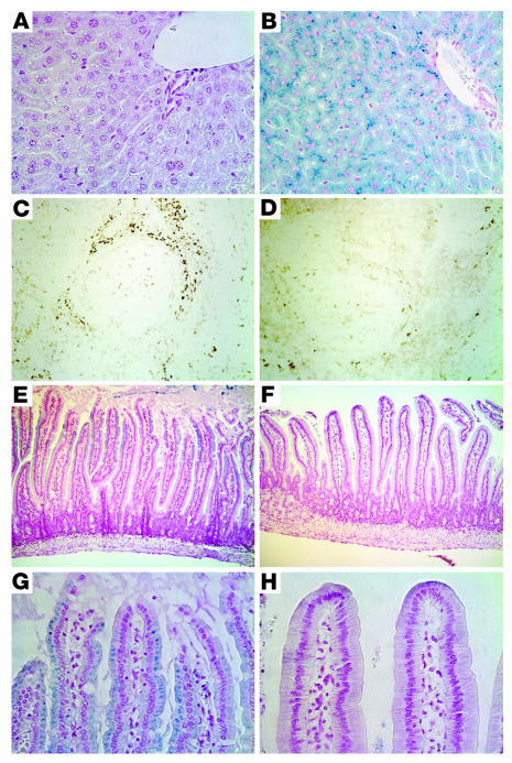
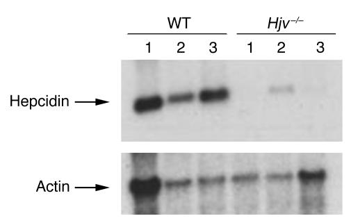
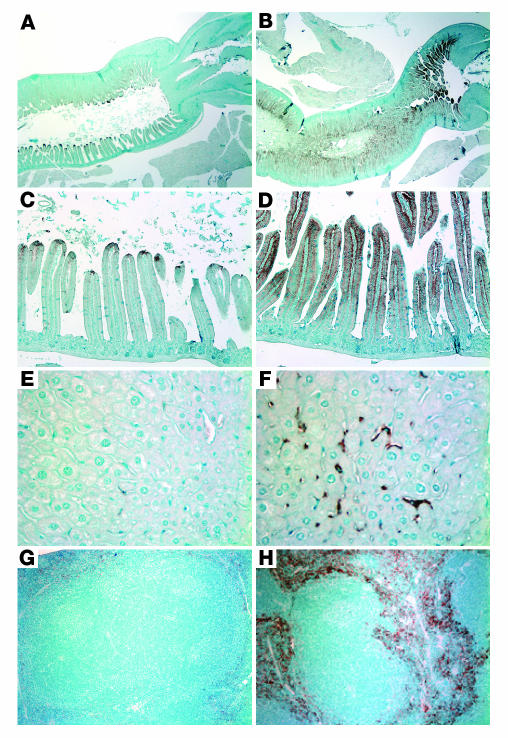
Hereditary hemochromatosis is an iron-overload disorder resulting from mutations in proteins presumed to be involved in the maintenance of iron homeostasis. Mutations in hemojuvelin (HJV) cause severe, early-onset juvenile hemochromatosis. The normal function of HJV is unknown. Juvenile hemochromatosis patients have decreased urinary levels of hepcidin, a peptide hormone that binds to the cellular iron exporter ferroportin, causing its internalization and degradation. We have disrupted the murine Hjv gene and shown that Hjv-/- mice have markedly increased iron deposition in liver, pancreas, and heart but decreased iron levels in tissue macrophages. Hepcidin mRNA expression was decreased in Hjv-/- mice. Accordingly, ferroportin expression detected by immunohistochemistry was markedly increased in both intestinal epithelial cells and macrophages. We propose that excess, unregulated ferroportin activity in these cell types leads to the increased intestinal iron absorption and plasma iron levels characteristic of the juvenile hemochromatosis phenotype.
Figures
Comment in
-
Of mice and men: the iron age.J Clin Invest. 2005 Aug;115(8):2079-82. doi: 10.1172/JCI25642. J Clin Invest. 2005. PMID: 16075054 Free PMC article.
References
-
- Hentze MW, Muckenthaler MU, Andrews NC. Balancing acts: molecular control of mammalian iron metabolism [review] Cell. 2004;117:285–297. - PubMed
-
- Camaschella C, et al. The gene TFR2 is mutated in a new type of haemochromatosis mapping to 7q22. Nat. Genet. 2000;25:14–15. - PubMed
-
- Feder JN, et al. A novel MHC class I-like gene is mutated in patients with hereditary haemochromatosis. Nat. Genet. 1996;13:399–408. - PubMed
-
- Njajou OT, et al. A mutation in SLC11A3 is associated with autosomal dominant hemochromatosis. Nat. Genet. 2001;28:213–214. - PubMed
Publication types
MeSH terms
Substances
Grants and funding
LinkOut - more resources
Full Text Sources
Other Literature Sources
Medical
Molecular Biology Databases
Research Materials
