. 2005 Oct 15;391(Pt 2):177-84.
doi: 10.1042/BJ20050885.

NuMA is a major acceptor of poly(ADP-ribosyl)ation by tankyrase 1 in mitosis

Affiliations

NuMA is a major acceptor of poly(ADP-ribosyl)ation by tankyrase 1 in mitosis

William Chang et al. Biochem J. .

Abstract

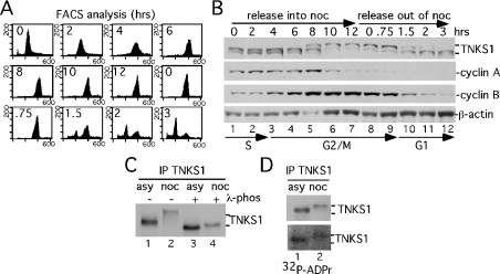
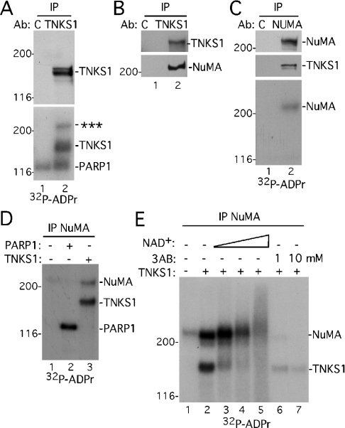
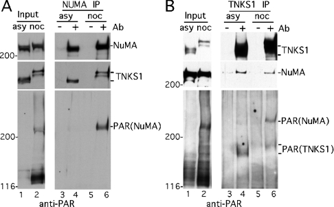
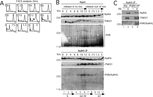
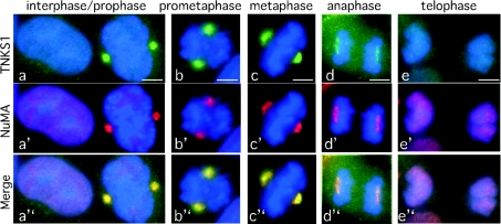
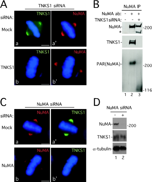
Tankyrase 1 is a PARP [poly(ADP-ribose) polymerase] that localizes to multiple subcellular sites, including telomeres and mitotic centrosomes. Previous studies demonstrated that cells deficient in tankyrase 1 suffered a block in resolution of sister telomeres and arrested in early anaphase [Dynek and Smith (2004) Science 304, 97-100]. This phenotype was dependent on the catalytic PARP activity of tankyrase 1. To identify critical acceptors of PARsylation [poly(ADP-ribosyl)ation] by tankyrase 1 in mitosis, tankyrase 1 immunoprecipitates were analysed for associated PARsylated proteins. We identified NuMA (nuclear mitotic apparatus protein) as a major acceptor of poly(ADP-ribose) from tankyrase 1 in mitosis. We showed by immunofluorescence and immunoprecipitation that association between tankyrase 1 and NuMA increases dramatically at the onset of mitosis, concomitant with PARsylation of NuMA. Knockdown of tankyrase 1 by siRNA (small interfering RNA) eliminates PARsylation of NuMA in mitosis, confirming tankyrase 1 as the PARP responsible for this modification. However, even in the absence of tankyrase 1 and PARsylation, NuMA localizes to spindle poles. By contrast, siRNA knockdown of NuMA results in complete loss of tankyrase 1 from spindle poles. We discuss our result in terms of a model where PARsylation of NuMA by tankyrase 1 in mitosis could play a role in sister telomere separation and/or mitotic progression.


Figures

Figure 1
Figure 1. Tankyrase 1 is modified in mitosis
(A, B) Analysis of staged cell cycle extracts shows a mitosis-specific mobility shift in tankyrase 1. Following arrest in S phase by a double thymidine block, HeLaI.2.11 cells were released into nocodazole for 12 h, collected by shake-off and released out of nocodazole for 3 h. (A) FACS analysis of staged extracts: y axis, cell numbers; x axis, relative DNA content based on propidium iodide staining. (B) Immunoblot analysis of staged cell extracts generated in buffer C. Products were detected with anti-tankyrase 1 antibody 465 (TNKS1), anti-cyclin A, anti-cyclin B or anti-β-actin antibodies. Cell cycle stages S, G2/M and G1 as determined by FACS (A) are indicated. (C) The tankyrase 1 mobility shift is due to phosphorylation. Extracts generated in TNE buffer from asynchronous (asy) or nocodazole (noc)-arrested cells were immunoprecipitated (IP) with anti-tankyrase 1 antibody 465, incubated with (+) or without (−) λ-phosphatase and products detected by immunoblotting with anti-tankyrase 1 465 (TNKS1). (D) Mitotic tankyrase 1 does not show an increase in autoPARsylation. Tankyrase 1 was immunoprecipitated as in (C) and incubated in a PARP assay with [32P]NAD+. Products were detected by immunoblotting with anti-tankyrase 1 antibody 465 (TNKS1; upper panel) or by autoradiography (32P-ADPr; lower panel).
Figure 2
Figure 2. A novel acceptor of PAR from tankyrase 1 in mitosis
(A) A novel acceptor of PAR co-immunoprecipitates with tankyrase 1. Extracts generated in TNE buffer from nocodazole-arrested HeLa.I.2.11 cells were immunoprecipitated (IP) with control IgG (C) or anti-tankyrase 1 antibody (Ab) 465 (TNKS1), and incubated in a PARP assay with [32P]NAD+. Products were detected by immunoblotting with anti-tankyrase 1 antibody 465 (TNKS1; upper panel) or by autoradiography (32P-ADPr; lower panel); *** indicates the novel acceptor. (B) NuMA co-immunoprecipitates with tankyrase 1. Extracts were immunoprecipitated as in (A) and products detected by immunoblotting with anti-tankyrase 1 antibody 465 or anti-NuMA. (C) NuMA is PARsylated. Extracts generated in TNE buffer from nocodazole-arrested cells were immunoprecipitated with control serum (C) or anti-NuMA and incubated in a PARP assay with [32P]NAD+. Products were detected by immunoblotting with anti-NuMA (top panel) or anti-tankyrase 1 antibody 465 (TNKS1; middle panel) or by autoradiography (32P-ADPr; bottom panel). (D) NuMA is PARsylated by recombinant tankyrase 1, not PARP1. NuMA was immunoprecipitated as in (C) and incubated in a PARP assay with [32P]NAD+ in the absence (−) or presence (+) of 2.5 μg of recombinant PARP1 or TNKS1. Products were detected by autoradiography. (E) PARsylation of NuMA is stimulated by NAD+ and inhibited by 3AB. NuMA was immunoprecipitated as in (C) and incubated in a PARP assay with [32P]NAD+ in the absence (−) or presence (+) of 2.5 μg of recombinant TNKS1 and increasing amounts of unlabelled NAD+ (0.04, 0.2 or 1 mM) or 3AB (1 or 10 mM). Products were detected by autoradiography.
Figure 3
Figure 3. NuMA is PARsylated in mitotic cells
(A) NuMA immunoprecipitated from mitotic cells is PARsylated. Extracts generated in buffer C from asynchronous (asy) or nocodazole (noc)-arrested HeLaI.2.11 cells were analysed directly (input) or immunoprecipitated (IP) with control serum (−) or anti-NuMA antibodies (Ab) (+). Products were detected by immunoblotting with anti-NuMA, anti-tankyrase 1 465 (TNKS1) or anti-PAR antibodies. (B) Tankyrase 1 immunoprecipitates from mitotic cells contain PARsylated NuMA. Extracts generated as in (A) were analysed directly (Input) or immunoprecipitated with control IgG (−) or anti-tankyrase 1 antibody 465 (+). Products were detected by immunoblotting with anti-tankyrase 1 465 (TNKS1), anti-NuMA or anti-PAR.
Figure 4
Figure 4. Tankyrase 1 associates with and PARsylates NuMA at the onset of mitosis
(A, B) Analysis of staged cell cycle extracts. Following arrest in S phase by a double thymidine block, HeLaI.2.11 cells were released into nocodazole for 12 h, collected by shake-off and released out of nocodazole for 3 h. (A) FACS analysis of staged extracts: y axis, cell numbers; x axis, relative DNA content based on propidium iodide staining. (B) Staged cell extracts generated in buffer C were analysed directly (Input) or immunoprecipitated (IP) with anti-NuMA. Products were detected by immunoblotting with anti-NuMA, anti-tankyrase 1 antibody 465 (TNKS1) or anti-PAR. Cell cycle stages S, G2/M and G1 as determined by FACS as in (A) are indicated. (C) PARsylation of NuMA does not depend on nocodazole. Extracts generated in buffer C from asynchronous (asy) or from mitotic shake-off 8–9 h after release from a double thymidine block (shake-off) were immunoprecipitated with control serum (−) or anti-NuMA antibodies (Ab) (+). Products were detected by immunoblotting with anti-NuMA, anti-tankyrase 1 antibody 465 (TNKS1) or anti-PAR.
Figure 5
Figure 5. Immunofluorescence analysis indicates co-localization of NuMA and tankyrase 1 from prophase through to anaphase
Cycling cells were methanol-fixed and stained with anti-tankyrase 1 antibody 609 (green) and anti-NuMA antibody 1F1 (red). DAPI staining of the DNA is blue. Co-localization is indicated as yellow in the merged images. Cell cycle stage is indicated at the top. Bar=5 μm.
Figure 6
Figure 6. Tankyrase 1 is required for PARsylation of NuMA, but not for localization of NuMA to spindle poles
(A) NuMA localizes to spindle poles in the absence of tankyrase 1. Immunofluorescence analysis of methanol-fixed HeLaI.2.11 cells is shown after 48 h of transfection with tankyrase 1 siRNA. Cells were stained with anti-tankyrase 1 antibody 609 (TNKS1; green) (a and b) or anti-NuMA 1F1 (red) (a′ and b′). DAPI staining of the DNA is in blue. Bar=5 μm. (B) siRNA of tankyrase 1 eliminates PARsylation of NuMA in mitosis. Cells were incubated with (+) or without (−) tankyrase 1 siRNA for 48 h and treated with nocodazole for 24 h prior to harvesting. Extracts generated in buffer C were immunoprecipitated (IP) with control serum (−) or anti-NuMA antibodies (ab) (+). Products were detected by immunoblotting with anti-NuMA (top panel), anti-tankyrase 1 antibody 465 (TNKS1; middle panel), or anti-PAR (bottom panel). The asterisk (*) indicates a NuMA breakdown product described previously [55,56]. (C) Knockdown of NuMA results in loss of tankyrase 1 at spindle poles. Immunofluorescence analysis of methanol-fixed HeLaI.2.11 cells is shown after 48 h of transfection with NuMA siRNA. Cells were stained with anti-NuMA antibody 1F1 (red) (a and b) or anti-tankyrase 1 antibody 609 (TNKS1; green) (a′ and b′). DAPI staining of the DNA is in blue. Bar=5 μm. (D) Immunoblot analysis of HeLa.I.2.11 cells mock transfected (−) or transfected with NuMA siRNA (+) for 48 h. Cell extracts were generated in buffer C and proteins were detected by immunoblotting with anti-NuMA, anti-tankyrase 1 465 (TNKS1) or anti-α-tubulin antibodies.

Comment in

Similar articles

Cited by

References

    1. Smith S., Giriat I., Schmitt A., de Lange T. Tankyrase, a poly(ADP-ribose) polymerase at human telomeres. Science. 1998;282:1484–1487. - PubMed
    1. Cook B. D., Dynek J. N., Chang W., Shostak G., Smith S. Role for the related poly(ADP-ribose) polymerases tankyrase 1 and 2 at human telomeres. Mol. Cell. Biol. 2002;22:332–342. - PMC - PubMed
    1. Smith S., de Lange T. Tankyrase promotes telomere elongation in human cells. Curr. Biol. 2000;10:1299–1302. - PubMed
    1. Dynek J. N., Smith S. Resolution of sister telomere association is required for progression through mitosis. Science. 2004;304:97–100. - PubMed
    1. de Lange T. Protection of mammalian telomeres. Oncogene. 2002;21:532–540. - PubMed

Publication types