. 2005 Aug 1;19(15):1811-22.
doi: 10.1101/gad.334805.

Control of replication initiation and heterochromatin formation in Saccharomyces cerevisiae by a regulator of meiotic gene expression

Affiliations

Control of replication initiation and heterochromatin formation in Saccharomyces cerevisiae by a regulator of meiotic gene expression

Horst Irlbacher et al. Genes Dev. .

Abstract

Heterochromatinization at the silent mating-type loci HMR and HML in Saccharomyces cerevisiae is achieved by targeting the Sir complex to these regions via a set of anchor proteins that bind to the silencers. Here, we have identified a novel heterochromatin-targeting factor for HML, the protein Sum1, a repressor of meiotic genes during vegetative growth. Sum1 bound both in vitro and in vivo to HML via a functional element within the HML-E silencer, and sum1Delta caused HML derepression. Significantly, Sum1 was also required for origin activity of HML-E, demonstrating a role of Sum1 in replication initiation. In a genome-wide search for Sum1-regulated origins, we identified a set of autonomous replicative sequences (ARS elements) that bound both the origin recognition complex and Sum1. Full initiation activity of these origins required Sum1, and their origin activity was decreased upon removal of the Sum1-binding site. Thus, Sum1 constitutes a novel global regulator of replication initiation in yeast.


Figures

Figure 1.
Figure 1.
Identification of a D-element core region within the HML-E silencer. (A) Schematic representation of the HMLα locus on the left arm of chromosome III. Location and elements of the silencers HML-E and HML-I are indicated. (RAP) Rap1-binding site; (ACS) ORC-binding site; (D) D element; (ABF) ABF-binding site. (B) Redundancy of HML-E silencer elements. Loss of HMLα silencing in HML-E silencer deletion mutants was measured as loss of a-mating ability in a patch mating assay. All strains were HML-ΔI. (C) D2 was the core element of D. Quantitative mating assays were performed to compare the effect on silencing of different D-element deletions in a MATa HML-E ACS- ΔI strain background (AEY3395). The mean values of at least three independent experiments are shown. (D) D2 was both necessary and sufficient for HML-E function. Loss of silencing in HML-E ACS- ΔI strains was measured for the 14-bp sequence element containing the D2 element (D, AEY3395), mutations in the entire (d2, AEY3426), the first (d2a, AEY3430) or the second (d2b, AEY3434) half of the D2 element, or with the D2 element remaining as the sole D sequence at HML-E (D2, AEY3552).
Figure 2.
Figure 2.
SUM1 was required for HMLα silencing and was epistatic to the D element. (A) Repression of HMLα or HMRa in strains deleted for SUM1, NAT1, or both was measured by patch mating assays. (B) SUM1, but not RFM1 or HST1, was genetically linked to HML-D. HMLα silencing of sum1Δ, rfm1Δ, or hst1Δ strains in combination with silencer element deletions at HML-E is shown by patch mating assays.
Figure 3.
Figure 3.
D-specific binding of Sum1 to HML-E in vitro and in vivo. (A) Sum1 bound in vitro to HML-E, but not the INO1 promoter region. (Left) A radioactively labeled 220-bp HML-E fragment was incubated without protein (lane 1) or with 0.1 μM bacterially expressed 6xHis-Sum1 (lanes 2-4). For competition experiments, unlabeled DNA of HML-E (specific competitor, lane 3) or a 210-bp INO1 fragment (unspecific competitor, lane 4) was added. DNA-protein complexes were resolved on a polyacrylamide gel and labeled DNA was visualized by autoradiography. (Right) Sum1 did not bind INO1 DNA, and bacterially expressed 6xHis-β-galactosidase (β-Gal) did not bind HML-E DNA. (Upper arrow) Protein-DNA complex. (Lower arrow) Free DNA. (B) Binding of Sum1 to HML-E required the D element. Mutant versions of HML-E were incubated with Sum1 (+) or without protein (-) and gel-electrophorezed as in A. HML-E DNA containing a mutation in the ACS site is termed ACS- (lanes 3,4,7,8), and HML-E DNA with deletion of the 93-bp D element is termed DΔ (lanes 5-8). To maintain DNA size in the DΔ derivates, the deleted D element was substituted for the genomic 3′-region of equivalent length. All DNA fragments were ∼220 bp. (C) Binding of Sum1 to HML-E required the D2 element. Mutant versions of HML-E were incubated with Sum1 (+) or without protein (-) as in A. (WT) A 134-bp wild-type HML-E fragment containing the ACS and the D element (lanes 1,2); (D2Δ) HML-E without the D2 element (lanes 3,4); (DΔ) a 140-bp HML-E fragment lacking the entire D element (lanes 5,6). (D) Competition between SMK1 and HML-E for Sum1 binding. A radioactively labeled double-stranded 19-bp fragment containing the MSE site of the SMK1 promoter was incubated without protein (lane 1) or with 0.1 μM bacterially expressed 6xHis-Sum1 (lanes 2-4). For competition experiments, unlabeled DNA of HML-E (specific competitor, lane 3) or HML-E DΔ was added. (E) Sum1 was associated in vivo with HML-E in a D-element-dependent manner. ChIPs were performed on sum1Δ strains containing a 2μ plasmid carrying N-terminally 6xmyc-tagged SUM1 under control of its own promoter (pAE1032). (Left) (WT) wild-type HMLα (AEY2); (ΔD) HMLΔDΔI (AEY3391). DNA was immunoprecipitated with (+) or without (-) anti-myc antibody and PCR-amplified. A total of 1/50 or 1/100 of the input DNA (lanes 7,8) or 1/2 (lanes 1,4), 1/4 (lanes 2,5), or 1/8 (lanes 3,6) of the immunoprecipitated DNA was analyzed. As a control, the promoter region of the SMK1 gene was PCR-amplified. (Right) ChIP was performed in sir4Δ strains. Columns indicate the ratio of DNA enrichment with versus without anti-myc antibody: (black columns) 6xmyc Sum1; (white columns) untagged. The Y-axis indicates fold enrichment.
Figure 4.
Figure 4.
Genetic interactions between SUM1 and replication initiation components. (A) sum1Δ reduced the ARS activity of HML-E. Plasmid loss rates were determined in a wild-type (AEY2) and a sum1Δ (AEY3358) strain. Strains with plasmids carrying ARS H4 (pRS316), HML-E (pAE1119), HMR-E (pAE229), or the HMR-E synthetic silencer SS HMR-E (pAE298) as their sole origins were analyzed. The average loss rates obtained from three independent experiments are shown with corresponding error bars. (B) Synthetic lethality of orc2-1 and sum1Δ. An orc2-1 sum1Δ strain carrying an URA3-labeled ORC2 plasmid (pRS316-ORC2) was transformed with a SUM1 (pAE1032) or an ORC2 (pAE53) plasmid or the corresponding empty vectors. Its ability to lose the pURA3-ORC2 plasmid was tested on 5-FOA medium. (C) Synthetic growth defects of cdc6-1, cdc7-1, or cdc45-1 with sum1Δ. Serial dilutions of several segregants from each cross were plated and incubated at the semipermissive temperature of the respective cdc single mutant. For cdc6-1, strains AEY600, 3358, and AEY3537 to 3541, for cdc7-1 strains AEY3542 to 3546, and for cdc45-1 stains AEY373 and AEY3548 to 3551 were used. Incubation was 3 d for cdc6-1 and cdc7-1 and 6 d for cdc45-1. cdc6-1 labeled with an asterisk indicates the parental strain, which was not isogenic to the sum1Δ strain. (D) Coimmunoprecipitation of Sum1 and Orc2. Strains AEY1558 (-) and AEY3474 (6xHis-Orc2, +) carried a 6xmyc-Sum1 2μ plasmid (pAE1032) and a HMLα (pAE1123) 2μ plasmid. Precipitates were analyzed by SDS-PAGE and immunoblotting using anti-myc-antibody. (Lanes 1,2) Input. (Lanes 3,4) Immunoprecipitation (IP). (Lanes 5,6) Supernatant (sup).
Figure 5.
Figure 5.
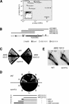
Sum1 was a replication initiation factor. (A) Plot of p-values for Sum1 binding (Lee et al. 2002), p < 0.01 versus ORC binding (Wyrick et al. 2001), p < 0.05. (Inset) All data points. The origin function of intergenic regions labeled with an asterisk was tested below. (B) Schematic representation of ARS1012 and ARS1013 located at the ORF YJL038C on chromosome X. The location of Ndt80 and Sum1 consensus sites (Pierce et al. 2003) and ACS matches is indicated. Bold lines represent fragments whose ARS function was tested. (C) SUM1 was required for ARS activity of ARS 1013 on plasmids. Strains AEY2 (WT) and AEY3358 (sum1Δ) were transformed with URA-CEN4 plasmids carrying either ARS1012 (pAE1076) or ARS1013-3 (pAE1081) as their sole origin. Transformants obtained upon transformation of ARS1013-1 or ARS1013-2-URA-CEN4 plasmids (pAE1078, pAE1080) were not restreakable. (D) SUM1 was required for chromosomal origin activity of ARS1013. The appearance of bubble-shaped replication intermediates indicative of chromosomal initiation (arrows) was measured by 2D gel electrophoresis and Southern hybridization in a wild-type (AEY2) and sum1Δ (AEY3358) strain. (E) Addition of Sum1-binding sites improved the ARS function of ARS1013-2. Strains AEY2 (WT) and AEY3358 (sum1Δ) were transformed with URA-CEN4 plasmids either carrying ARS1013-3 (pAE1081) or variants of ARS1013-2 containing additional fragments of HML-E (4xD2, pAE1159 and HML-E ACS-, pAE1160) or the SMK1 promoter (pAE1161) upstream of the ARS1013-2 fragment.
Figure 6.
Figure 6.
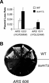
Sum1 affected ARS activity of selected origins of replication. (A) ARS1223 and ARS1511 required SUM1 for full ARS activity. Plasmid loss rates were determined in a wild-type (WT; AEY2) and a sum1Δ (AEY3358) strain. Strains with URA-CEN4 plasmids carrying ARS1223 (pAE1130) or ARS1511 (pAE1135) as their sole origins were analyzed. The average loss rates obtained from three independent experiments are shown with corresponding error bars. The loss rate in sum1Δ strains was approximately twofold (ARS1223) and 5.7-fold (ARS1511) higher than in wild-type strains. (B) ARS activity of ARS606 was dependent on SUM1. Strains AEY2 (WT) and AEY3358 (sum1Δ) were transformed with URA-CEN4 plasmids carrying ARS606 (pAE1126) as their sole origin and streaked on a -Ura plate.

Similar articles

Cited by

References

    1. Aparicio J.G., Viggiani, C.J., Gibson, D.G., and Aparicio, O.M. 2004. The Rpd3-Sin3 histone deacetylase regulates replication timing and enables intra-S origin control in Saccharomyces cerevisiae. Mol. Cell. Biol. 24: 4769-4780. - PMC - PubMed
    1. Beall E.L., Manak, J.R., Zhou, S., Bell, M., Lipsick, J.S., and Botchan, M.R. 2002. Role for a Drosophila Myb-containing protein complex in site-specific DNA replication. Nature 420: 833-837. - PubMed
    1. Bell S.P. and Dutta, A. 2002. DNA replication in eukaryotic cells. Annu. Rev. Biochem. 71: 333-374. - PubMed
    1. Bell S.P., Kobayashi, R., and Stillman, B. 1993. Yeast origin recognition complex functions in transcription silencing and DNA replication. Science 262: 1844-1849. - PubMed
    1. Breier A.M., Chatterji, S., and Cozzarelli, N.R. 2004. Prediction of Saccharomyces cerevisiae replication origins. Genome Biol. 5: R22. - PMC - PubMed

Publication types