. 2005 Aug;187(16):5560-7.
doi: 10.1128/JB.187.16.5560-5567.2005.

Upstream migration of Xylella fastidiosa via pilus-driven twitching motility

Affiliations

Upstream migration of Xylella fastidiosa via pilus-driven twitching motility

Yizhi Meng et al. J Bacteriol. 2005 Aug.

Abstract

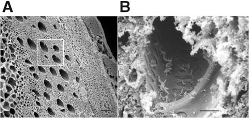
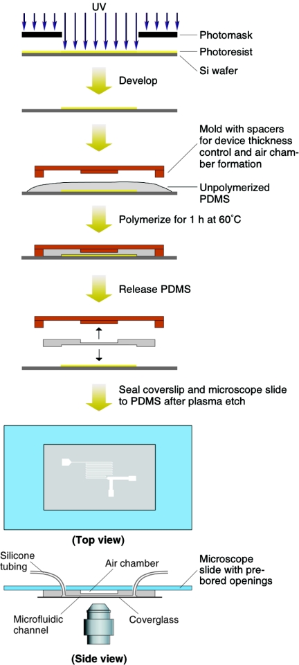
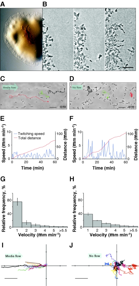
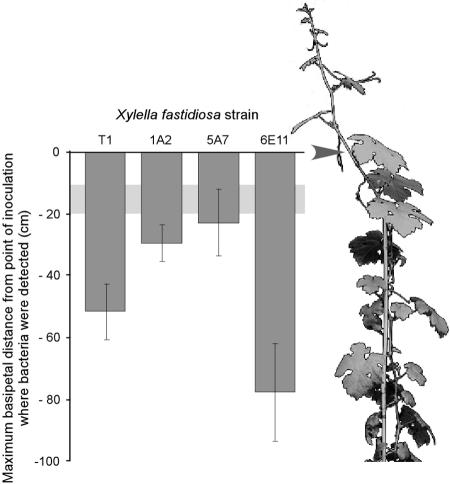
Xylella fastidiosa is a xylem-limited nonflagellated bacterium that causes economically important diseases of plants by developing biofilms that block xylem sap flow. How the bacterium is translocated downward in the host plant's vascular system against the direction of the transpiration stream has long been a puzzling phenomenon. Using microfabricated chambers designed to mimic some of the features of xylem vessels, we discovered that X. fastidiosa migrates via type IV-pilus-mediated twitching motility at speeds up to 5 mum min(-1) against a rapidly flowing medium (20,000 mum min(-1)). Electron microscopy revealed that there are two length classes of pili, long type IV pili (1.0 to 5.8 mum) and short type I pili (0.4 to 1.0 mum). We further demonstrated that two knockout mutants (pilB and pilQ mutants) that are deficient in type IV pili do not twitch and are inhibited from colonizing upstream vascular regions in planta. In addition, mutants with insertions in pilB or pilQ (possessing type I pili only) express enhanced biofilm formation, whereas a mutant with an insertion in fimA (possessing only type IV pili) is biofilm deficient.


Figures

FIG. 1.
FIG. 1.
Scanning electron micrographs of tangential sections through grapevine and associated xylem vessels. (A) Water-conducting xylem vessels (several of which are noted within the white square) of a grapevine cross section serve as the habitat for X. fastidiosa cells. Bar, 200 μm. (B) View into a sectioned xylem vessel with X. fastidiosa cells attached to the vessel walls. Bar, 10 μm.
FIG. 2.
FIG. 2.
Fabrication process and mounting of the microfluidic chamber (100 μm deep, 100 μm wide, and 14 cm long). Si, silicon.
FIG. 3.
FIG. 3.
Twitching motility characteristics of wild-type X. fastidiosa. (A) Light micrograph of a colony with a peripheral fringe. (B) Sequential images (0 min, 7 h 18 min, and 14 h 20 min, left to right) depicting progressive migration of twitching cells (see movie 1A in the supplemental material; also see expanded versions and additional movies at http://www.nysaes.cornell.edu/pp/faculty/hoch/movies/). Bar, 20 μm. (C) Light micrograph depicting paths of three bacteria (circled) twitching against a flow of 20,000 μm min−1 (see movie 2 in the supplemental material and the expanded versions and additional movies at the above-cited website). Bar, 10 μm. (D) Similar depiction as in panel C, but with no flow (see movie 3 in the supplemental material and the expanded versions and additional movies at the above-cited website). Bar, 10 μm. (E and F) Twitching speed and cumulative distance traveled by the red-circled cells in panels C and D, respectively. (G and H) Histograms depicting twitching speeds for cells under flow (20,000 μm min−1) (G) and no-flow (H) conditions. Error bars represent standard errors of the mean (n = 17). (I and J) Twitching paths (starting at coordinates 0,0) measured in a 1-h period for cells under conditions of flow (20,000 μm min−1) (I) and no flow (J). Bar, 20 μm.
FIG. 4.
FIG. 4.
Pili of wild-type and mutant X. fastidiosa. (A) Scanning electron micrograph of wild-type cells attached to the substratum at the pilus-bearing polar ends. Many pili degenerated into globular masses during specimen preparation. (B and C) Transmission electron micrographs of negatively stained (phosphotungstic acid) wild-type cells depicting an abundance of short pili (B), and fewer long type IV pili (C). (D and E) Negatively stained preparations of mutants 1A2 and 6E11 depicting only short pili and only longer (type IV) pili, respectively. Pilus lengths are noted in Fig. 8. Bars, 1 μm.
FIG. 5.
FIG. 5.
Xylella fastidiosa mutant characteristics. (A) Light micrograph of X. fastidiosa mutant 1A2 colony on agar medium with a well-demarcated fringeless periphery. (B) Sequential images (0 min and 20 h 19 min, respectively) depicting progressive expansion of the 1A2 colony (see movie 1B in the supplemental material; also see expanded versions and additional movies at http://www.nysaes.cornell.edu/pp/faculty/hoch/movies/). Bar, 20 μm.
FIG. 6.
FIG. 6.
Genomic organization of genes flanking Tn5 insertions in X. fastidiosa mutants 1A2, 5A7, and 6E11. nt, nucleotide.
FIG. 7.
FIG. 7.
Biofilm production and growth curves of the X. fastidiosa wild type and mutants. (A) Biofilm formation by the X. fastidiosa wild type and mutants 1A2, 5A7, and 6E11 following 7-day growth at 28°C. Biofilms were stained with 0.1% crystal violet. (B) The crystal violet stain was subsequently removed from the tubes with dimethyl sulfoxide and quantified for optical density at 620 nm. OD, optical density. (C) Growth curves for the X. fastidiosa wild type and mutants 1A2, 5A7, and 6E11 in PW medium at 28°C.
FIG. 8.
FIG. 8.
Pilus measurement in X. fastidiosa. Histograms of pilus length distributions for the wild type (A) and mutants 1A2 (B) and 6E11 (C). Number of cells represented were 11 (A), 12 (B), and 14 (C).
FIG. 9.
FIG. 9.
Basipetal translocation of X. fastidiosa in planta. Maximum distances for wild-type and mutant X. fastidiosa cells were recovered from grapevine regions upstream of the inoculation sites (represented by 0 on the y axis; arrow of illustrated vine) after 11 weeks. The light-gray horizontal band is the zone representing the maximum distance that 0.2-μm fluorescent latex beads traveled passively. Error bars represent standard errors of the means.

References

    1. Alm, R. A., and J. S. Mattick. 1997. Genes involved in the biogenesis and function of type-4 fimbriae in Pseudomonas aeruginosa. Gene 192:89-98. - PubMed
    1. Bradley, D. E. 1980. A function of Pseudomonas aeruginosa PAO polar pili: twitching motility. Can. J. Microbiol. 26:146-154. - PubMed
    1. Chaudhury, M. K., and G. M. Whitesides. 1991. Direct measurement of interfacial interactions between semispherical lenses and flat sheets of poly(dimethylsiloxane) and their chemical derivatives. Langmuir 7:1013-1025.
    1. Davis, M. J., W. J. French, and N. W. Schaad. 1981. Axenic culture of the bacteria associated with phony disease of peach and plum leaf scald. Curr. Microbiol. 6:309-314.
    1. Feil, H., W. S. Feil, J. C. Detter, A. H. Purcell, and S. E. Lindow. 2003. Site-directed disruption of the fimA and fimF genes of Xylella fastidiosa. Phytopathology 93:675-682. - PubMed

Publication types

LinkOut - more resources