Biochemical characterization of RssA-RssB, a two-component signal transduction system regulating swarming behavior in Serratia marcescens
- PMID: 16077114
- PMCID: PMC1196059
- DOI: 10.1128/JB.187.16.5683-5690.2005
Biochemical characterization of RssA-RssB, a two-component signal transduction system regulating swarming behavior in Serratia marcescens
Abstract
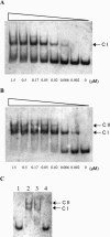
Our previous study had identified a pair of potential two-component signal transduction proteins, RssA-RssB, involved in the regulation of Serratia marcescens swarming. When mutated, both rssA and rssB mutants showed precocious swarming phenotypes on LB swarming agar, whereby swarming not only occurred at 37 degrees C but also initiated on a surface of higher agar concentration and more rapidly than did the parent strain at 30 degrees C. In this study, we further show that the predicted sensor kinase RssA and the response regulator RssB bear characteristics of components of the phosphorelay signaling system. In vitro phosphorylation and site-directed mutagenesis assays showed that phosphorylated RssA transfers the phosphate group to RssB and that histidine 248 and aspartate 51 are essential amino acid residues involved in the phosphotransfer reactions in RssA and RssB, respectively. Accordingly, while wild-type rssA could, the mutated rssA(H248A) in trans could not complement the precocious swarming phenotype of the rssA mutant. Although RssA-RssB regulates expressions of shlA and ygfF of S. marcescens (ygfF(Sm)), in vitro DNA-binding assays showed that the phosphorylated RssB did not bind directly to the promoter regions of these two genes but bound to its own rssB promoter. Subsequent assays located the RssB binding site within a 63-bp rssB promoter DNA region and confirmed a direct negative autoregulation of the RssA-RssB signaling pathway. These results suggest that when activated, RssA-RssB acts as a negative regulator for controlling the initiation of S. marcescens swarming.
Figures


Similar articles
-
The RssAB two-component signal transduction system in Serratia marcescens regulates swarming motility and cell envelope architecture in response to exogenous saturated fatty acids.J Bacteriol. 2005 May;187(10):3407-14. doi: 10.1128/JB.187.10.3407-3414.2005. J Bacteriol. 2005. PMID: 15866926 Free PMC article.
-
Regulation of swarming motility and flhDC(Sm) expression by RssAB signaling in Serratia marcescens.J Bacteriol. 2008 Apr;190(7):2496-504. doi: 10.1128/JB.01670-07. Epub 2008 Jan 25. J Bacteriol. 2008. PMID: 18223092 Free PMC article.
-
ManA is regulated by RssAB signaling and promotes motility in Serratia marcescens.Res Microbiol. 2014 Jan;165(1):21-9. doi: 10.1016/j.resmic.2013.10.005. Epub 2013 Oct 23. Res Microbiol. 2014. PMID: 24161484
-
Activation and secretion of Serratia hemolysin.Zentralbl Bakteriol. 1993 Apr;278(2-3):306-15. doi: 10.1016/s0934-8840(11)80847-9. Zentralbl Bakteriol. 1993. PMID: 8347934 Review.
-
A signal peptide-independent protein secretion pathway.Antonie Van Leeuwenhoek. 1992 Feb;61(2):111-3. doi: 10.1007/BF00580616. Antonie Van Leeuwenhoek. 1992. PMID: 1580612 Review. No abstract available.
Cited by
-
Proteomic profiling of Serratia marcescens by high-resolution mass spectrometry.Bioimpacts. 2020;10(2):123-135. doi: 10.34172/bi.2020.15. Epub 2020 Mar 26. Bioimpacts. 2020. PMID: 32363156 Free PMC article.
-
An iron detection system determines bacterial swarming initiation and biofilm formation.Sci Rep. 2016 Nov 15;6:36747. doi: 10.1038/srep36747. Sci Rep. 2016. PMID: 27845335 Free PMC article.
-
RssAB signaling coordinates early development of surface multicellularity in Serratia marcescens.PLoS One. 2011;6(8):e24154. doi: 10.1371/journal.pone.0024154. Epub 2011 Aug 26. PLoS One. 2011. PMID: 21887380 Free PMC article.
-
RssAB-FlhDC-ShlBA as a major pathogenesis pathway in Serratia marcescens.Infect Immun. 2010 Nov;78(11):4870-81. doi: 10.1128/IAI.00661-10. Epub 2010 Aug 16. Infect Immun. 2010. PMID: 20713626 Free PMC article.
-
A mobile quorum-sensing system in Serratia marcescens.J Bacteriol. 2006 Feb;188(4):1518-25. doi: 10.1128/JB.188.4.1518-1525.2006. J Bacteriol. 2006. PMID: 16452435 Free PMC article.
References
-
- Bird, T. H., S. Du, and C. E. Bauer. 1999. Autophosphorylation, phosphotransfer, and DNA-binding properties of the RegB/RegA two-component regulatory system in Rhodobacter capsulatus. J. Biol. Chem. 274:16343-16348. - PubMed
-
- Cserzo, M., E. Wallin, I. Simon, G. von Heijne, and A. Elofsson. 1997. Prediction of transmembrane alpha-helices in prokaryotic membrane proteins: the dense alignment surface method. Protein Eng. 10:673-676. - PubMed
-
- Cybulski, L. E., G. del Solar, P. O. Craig, M. Espinosa, and D. de Mendoza. 2004. Bacillus subtilis DesR functions as a phosphorylation-activated switch to control membrane lipid fluidity. J. Biol. Chem. 279:39340-39347. - PubMed
Publication types
MeSH terms
Substances
Associated data
- Actions
LinkOut - more resources
Full Text Sources

