

Combustion-derived ultrafine particles transport organic toxicants to target respiratory cells
- PMID: 16079063
- PMCID: PMC1280333
- DOI: 10.1289/ehp.7661
Combustion-derived ultrafine particles transport organic toxicants to target respiratory cells
Abstract
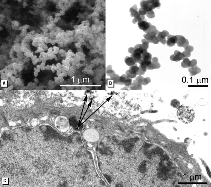
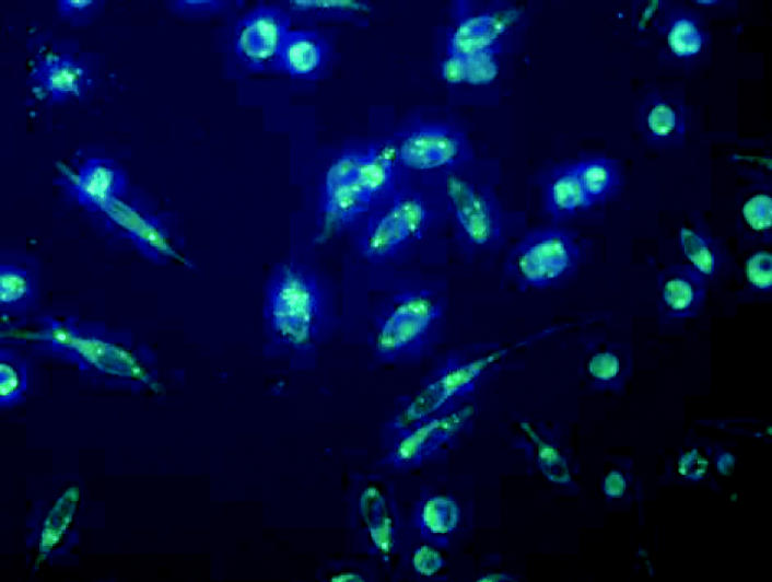
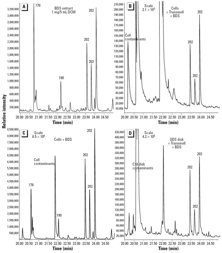
Epidemiologic evidence supports associations between inhalation of fine and ultrafine ambient particulate matter [aerodynamic diameter < or = 2.5 microm (PM2.5)] and increases in cardiovascular/respiratory morbidity and mortality. Less attention has been paid to how the physical and chemical characteristics of these particles may influence their interactions with target cells. Butadiene soot (BDS), produced during combustion of the high-volume petrochemical 1,3-butadiene, is rich in polynuclear aromatic hydrocarbons (PAHs), including known carcinogens. We conducted experiments to characterize BDS with respect to particle size distribution, assembly, PAH composition, elemental content, and interaction with respiratory epithelial cells. Freshly generated, intact BDS is primarily (> 90%) PAH-rich, metals-poor (nickel, chromium, and vanadium concentrations all < 1 ppm) PM2.5, composed of uniformly sized, solid spheres (30-50 nm) in aggregated form. Cells of a human bronchial epithelial cell line (BEAS-2B) exhibit sequential fluorescent responses--a relatively rapid (approximately 30 min), bright but diffuse fluorescence followed by the slower (2-4 hr) appearance of punctate cytoplasmic fluorescence--after BDS is added to medium overlying the cells. The fluorescence is associated with PAH localization in the cells. The ultrafine BDS particles move down through the medium to the cell membrane. Fluorescent PAHs are transferred from the particle surface to the cell membrane, cross the membrane into the cytosol, and appear to accumulate in lipid vesicles. There is no evidence that BDS particles pass into the cells. The results demonstrate that uptake of airborne ultrafine particles by target cells is not necessary for transfer of toxicants from the particles to the cells.
Figures
References
-
- Allison AC, Mallucci L. Uptake of hydrocarbon carcinogens by lysosomes. Nature. 1964;203:1024–1027. - PubMed
-
- Bermudez E, Mangum JB, Wong BA, Ashgarian B, Hext PM, Warheit DB, et al. Pulmonary responses of mice, rats and hamsters to subchronic inhalation of ultrafine titanium dioxide particles. Toxicol Sci. 2004;77:347–357. - PubMed
-
- Berube KA, Jones TP, Williamson BJ, Winters C, Morgan AJ, Richards JR. Physiochemical characterization of diesel exhaust particles: factors for assessing biological activity. Atmos Environ. 1999;33:1599–1614.
-
- Boland S, Baeza-Squiban A, Fournier T, Houcine O, Gendron MC, Chévrier M, et al. Diesel exhaust particles are taken up by human airway epithelial cells in vitro and alter cytokine production. Am J Physiol Lung Cell Mol Physiol. 1999;276:L604–L613. - PubMed
-
- Bonvallot V, Baeza-Squiban A, Baulig A, Brulant S, Boland S, Muzeau F, et al. Organic compounds from diesel exhaust particles elicit a proinflammatory response in human airway epithelial cells and induce cytochrome p450 1A1 expression. Am J Respir Cell Mol Biol. 2001;25:515–521. - PubMed
Publication types
MeSH terms
Substances
LinkOut - more resources
Full Text Sources