. 2005 Oct;16(10):4827-40.
doi: 10.1091/mbc.e05-04-0315. Epub 2005 Aug 3.

Active Nercc1 protein kinase concentrates at centrosomes early in mitosis and is necessary for proper spindle assembly

Affiliations

Active Nercc1 protein kinase concentrates at centrosomes early in mitosis and is necessary for proper spindle assembly

Joan Roig et al. Mol Biol Cell. 2005 Oct.

Abstract

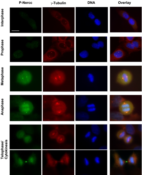
The Nercc1 protein kinase autoactivates in vitro and is activated in vivo during mitosis. Autoactivation in vitro requires phosphorylation of the activation loop at threonine 210. Mitotic activation of Nercc1 in mammalian cells is accompanied by Thr210 phosphorylation and involves a small fraction of total Nercc1. Mammalian Nercc1 coimmunoprecipitates gamma-tubulin and the activated Nercc1 polypeptides localize to the centrosomes and spindle poles during early mitosis, suggesting that active Nercc has important functions at the microtubular organizing center during cell division. To test this hypothesis, we characterized the Xenopus Nercc1 orthologue (XNercc). XNercc endogenous to meiotic egg extracts coprecipitates a multiprotein complex that contains gamma-tubulin and several components of the gamma-tubulin ring complex and localizes to the poles of spindles formed in vitro. Reciprocally, immunoprecipitates of the gamma-tubulin ring complex polypeptide Xgrip109 contain XNercc. Immunodepletion of XNercc from egg extracts results in delayed spindle assembly, fewer bipolar spindles, and the appearance of aberrant microtubule structures, aberrations corrected by addition of purified recombinant XNercc. XNercc immunodepletion also slows aster assembly induced by Ran-GTP, producing Ran-asters of abnormal size and morphology. Thus, Nercc1 contributes to both the centrosomal and the chromatin/Ran pathways that collaborate in the organization of a bipolar spindle.


Figures

Figure 1.
Figure 1.
Phosphorylation of Nercc1 kinase (Thr210) during autoactivation in vitro and during mitosis. (A) Autoactivation of recombinant Nercc1 in vitro requires Thr210. Wild-type and T210A mutant forms of Nercc1 were expressed in HEK293T cells, immunoprecipitated using FLAG antibody, washed, and incubated for the indicated times with phosphorylation buffer containing [γ-32P]ATP and model substrate histone H3. FLAG Western blots, and 32P autoradiographs of XNercc and histone H3 are shown. (B) Specificity of an anti-phospho-Thr210-Nercc1 antibody. Recombinant FLAG-Nercc1 was expressed in HEK293T cells, immunoprecipitated using FLAG antibody, washed, and incubated in phosphorylation buffer with or without 100 μM ATP. Western blots using FLAG antibody and purified anti-phospho-Thr210-Nercc1 (a-P-Nercc1) antibody are shown. (C) Anti-Nercc1 and anti-phospho-Thr210-Nercc1 immunoblot of anti-Nercc1 polypeptide immunoprecipitates. Immunoprecipitates prepared from extracts of exponentially growing (Exp) or nocodazole arrested, mitotic (M) U2OS cells using normal rabbit IgG (NIgG, lanes 1, 2, 7, and 8) or anti-total Nercc1 antibody raised against the N-terminal (a-Nercc1 [Nt], lanes 3, 4, 9, and 10) or the C-terminal tail (a-Nercc1 [Ct], lanes 5, 6, 11, and 12) of Nercc1, were subjected to immunoblot with anti-Nercc1 (Nt) polypeptide (lanes 1-6) and anti-phospho-Thr210-Nercc1 (lanes 7-12). (D) Anti-phospho Thr210 Nercc1 immunoblot of anti-Nercc1 polypeptide immunoprecipitates. Immunoprecipitates from extracts of exponentially growing (Exp) or mitotic (M) U2OS cells are immunoblotted for Nercc1 polypeptide (lanes 1-4 and 9-12) or phospho-Thr210-Nercc1 (lanes 5-8). An overexposure of the blot in lanes 1-4 is shown in lanes 9-12 to make visible the high-molecular weight form of Nercc1, indicated by the asterisk, that is present in mitotic cells.
Figure 2.
Figure 2.
Activated Nercc1 is concentrated at the centrosome and binds γ-tubulin. (A) Specificity of the anti-Nercc1 Thr210P immunoblot. An anti-phospho-Thr210-Nercc1 immunoblot of total cell extracts from exponentially growing (Exp) and mitotic U2OS cells (M, arrested by overnight incubation with nocodazole). (B) Immunolocalization of active Nercc1 in interphase U2OS cells using the anti-phospho-Thr210 Nercc1 antibodies. Costaining is with anti-γ-tubulin and DAPI (DNA). Bar, 15 μm. (C) P-peptide competition of anti-phospho-Thr210 Nercc1 immunofluorescence. As in A, but the antibodies were preincubated either with no peptide (top row) or the immunizing peptide in its unphosphorylated (middle row) or phosphorylated (bottom row) form. For clarity, prometaphase cells are shown. Bar, 15 μm. (D) Nercc1 coprecipitatesγ-tubulin. Immunoprecipitates prepared from extracts of exponentially growing (Exp) or nocodazole arrested, mitotic (M) U2OS cells using normal rabbit IgG (NIgG), or anti-total Nercc1 antibody (a-Nercc1) were subjected to immunoblot with either anti-Nercc1 (top) or anti-γ-tubulin (bottom) antibodies.
Figure 3.
Figure 3.
Immunolocalization of active Nercc1 during mitotic progression in U2OS cells. Staining of active Nercc1, DNA, and γ-tubulin as described in A. Bar, 15 μm.
Figure 4.
Figure 4.
Identification and characterization of the X. laevis Nercc1 orthologue, XNercc. (A) Protein sequence alignment of X. laevis Nercc1 (XNercc) versus human Nercc1 (hNercc). (B) Recombinant XNercc autoactivates in vitro. FLAG-XNercc immunoprecipitated from transfected HEK293 cells was incubated with phosphorylation buffer containing [γ-32P]ATP(100 μM) and the model substrate MBP. At the times indicated, aliquots were removed, subjected to SDS-PAGE. Coomassie stain of the FLAG-XNercc (top) and 32P autoradiography of the FLAG-XNercc (middle) and MBP (bottom) are shown. (C) Immunoblot of a Xenopus mitotic egg extract for XNercc polypeptide. The affinity purified anti-XNercc antibody was used. (D) Identification of Xenopus egg proteins that coprecipitate with endogenous XNercc. Immunoprecipitates prepared from mitotic Xenopus egg extracts using normal rabbit IgG (NIgG) or anti-XNercc (a-XNercc) were washed repeatedly and subjected to SDS-PAGE. After Coomassie staining, the numbered bands were excised, digested with trypsin, and analyzed by LC/MS/MS; the polypeptides identified in each band were 1, C6/Xgrip210; 2, XNercc; 3, major vault protein; 4, GCP3/Xgrip110 and GCP2/Xgrip109; 5, HSP70; and 6, γ-tubulin. (E) XNercc coprecipitates with the γTuRC. Left, immunoprecipitates prepared from mitotic egg extracts using normal IgG (NIgG) or anti-XNercc were subjected to immunoblot using anti-XNercc (top) and anti-γ-tubulin (bottom). Right, Xgrip109 immunoprecipitates (top) contain XNercc (middle) and γ-tubulin (bottom).
Figure 5.
Figure 5.
Immunolocalization of XNercc in mitotic spindles assembled in vitro from Xenopus egg extracts. Mitotic spindles were assembled in vitro from Xenopus egg extracts as described in Materials and Methods. Aliquots were stained either with anti-XNercc antibody (bottom) or normal IgG (NIgG, top). Microtubules are visualized with rhodamine-labeled tubulin and DNA with DAPI. A magnified example of a bipolar spindle showing XNercc polypeptide localization to the poles is shown (bottom). Bar, 10 μm.
Figure 6.
Figure 6.
XNercc immunodepletion from mitotic egg extracts causes delayed and aberrant spindle assembly. (A) Anti-XNercc immunoblot of Xenopus egg extracts after three cycles of immunoprecipitation with either normal IgG or two different anti-XNercc antibodies. After three rounds of immunoprecipitation with normal IgG (NIgG), C-XNercc antibody (C-X), and N-XNercc antibody (N-X), the residual extract was subjected to immunoblot with N-XNercc antibody. Right, immunoblot for γ-tubulin of extracts subjected to immunodepletion using normal IgG or anti-N-XNercc antibodies. (B) Representative spindle structures formed in control and XNercc-depleted extracts. Mitotic egg extracts were immunodepleted using the three IgG preparations described in A. After addition of demembranated sperm nuclei, the extracts where cycled once by calcium addition. Monopoles and spindles were visualized with rhodamine-labeled tubulin and DAPI after 40 min. Control ID, extract after three round of immunoprecipitation with normal rabbit IgG; N-XNercc ID, extract after three round of immunoprecipitation with N-XNercc antibody. Bar, 10 μm. (C) Addition of purified recombinant XNercc corrects the defective spindle assembly of XNercc-depleted egg extracts. The results of two experiments are shown; spindles were enumerated at 60-75 min after addition of sperm nuclei. AB, XNercc add back before sperm nuclei; other, multipolar structures.
Figure 7.
Figure 7.
XNercc depletion interferes with RanQ69L(GTP)-induced aster formation. Xenopus egg extracts were subjected to three rounds of immunoprecipitation with normal rabbit IgG (Control ID) or N-XNercc antibody (N-XNercc ID) as shown in Figure 6. At several times after addition of GTP-loaded RanQ69L rhodamine-labeled tubulin structures were quantified at low magnification. A representative time course of aster formation on the immunodepleted extracts is shown in A. (B) Structures formed in mock or XNercc-depleted extracts after 20 min of GTP-loaded RanQ69L addition. XNercc depletion, in addition to causing a delay in the appearance of tubulin assemblies, results in the appearance of smaller, more condensed structures that tend to cluster (B). Bar, 10 μm.

Similar articles

Cited by

References

    1. Belham, C., Roig, J., Caldwell, J. A., Aoyama, Y., Kemp, B. E., Comb, M., and Avruch, J. (2003). A mitotic cascade of NIMA family kinases. Nercc1/Nek9 activates the Nek6 and Nek7 kinases. J. Biol. Chem. 278, 34897-34909. - PubMed
    1. Bischoff, F. R., Klebe, C., Kretschmer, J., Wittinghofer, A., and Ponstingl, H. (1994). RanGAP1 induces GTPase activity of nuclear Ras-related Ran. Proc. Natl. Acad. Sci. USA 91, 2587-2591. - PMC - PubMed
    1. Carazo-Salas, R. E., Guarguaglini, G., Gruss, O. J., Segref, A., Karsenti, E., and Mattaj, I. W. (1999). Generation of GTP-bound Ran by RCC1 is required for chromatin-induced mitotic spindle formation. Nature 400, 178-181. - PubMed
    1. Desai, A., Murray, A., Mitchison, T. J., and Walczak, C. E. (1999). The use of Xenopus egg extracts to study mitotic spindle assembly and function in vitro. Methods Cell Biol. 61, 385-412. - PubMed
    1. Doxsey, S. (2001). Re-evaluating centrosome function. Nat. Rev. Mol. Cell. Biol. 2, 688-698. - PubMed

Publication types

LinkOut - more resources