Comparative Study
. 2005 Oct;16(10):4714-24.
doi: 10.1091/mbc.e05-03-0246. Epub 2005 Aug 3.

Characterization of Schizosaccharomyces pombe ER alpha-mannosidase: a reevaluation of the role of the enzyme on ER-associated degradation

Affiliations
Comparative Study

Characterization of Schizosaccharomyces pombe ER alpha-mannosidase: a reevaluation of the role of the enzyme on ER-associated degradation

Federico Movsichoff et al. Mol Biol Cell. 2005 Oct.

Abstract

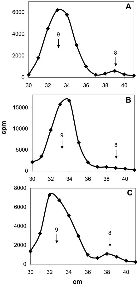
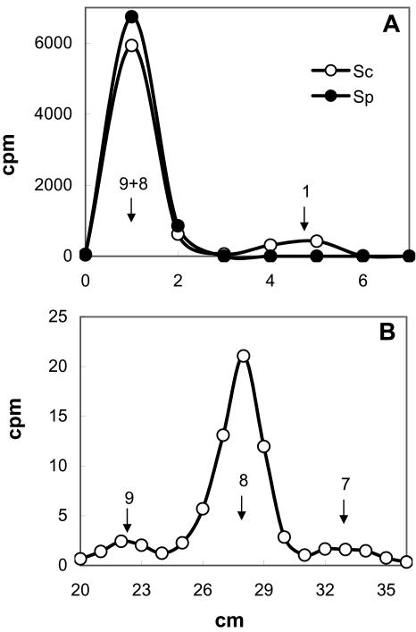
It has been postulated that creation of Man8GlcNAc2 isomer B (M8B) by endoplasmic reticulum (ER) alpha-mannosidase I constitutes a signal for driving irreparably misfolded glycoproteins to proteasomal degradation. Contrary to a previous report, we were able to detect in vivo (but not in vitro) an extremely feeble ER alpha-mannosidase activity in Schizosaccharomyces pombe. The enzyme yielded M8B on degradation of Man9GlcNAc2 and was inhibited by kifunensin. Live S. pombe cells showed an extremely limited capacity to demannosylate Man9GlcNAc2 present in misfolded glycoproteins even after a long residence in the ER. In addition, no preferential degradation of M8B-bearing species was detected. Nevertheless, disruption of the alpha-mannosidase encoding gene almost totally prevented degradation of a misfolded glycoprotein. This and other conflicting reports may be best explained by assuming that the role of ER mannosidase on glycoprotein degradation is independent of its enzymatic activity. The enzyme, behaving as a lectin binding polymannose glycans of varied structures, would belong together with its enzymatically inactive homologue Htm1p/Mnl1p/EDEM, to a transport chain responsible for delivering irreparably misfolded glycoproteins to proteasomes. Kifunensin and 1-deoxymannojirimycin, being mannose homologues, would behave as inhibitors of the ER mannosidase or/and Htm1p/Mnl1p/EDEM putative lectin properties.


Figures

Figure 1.
Figure 1.
Glycan structures. The figure represents the glycan transferred to proteins in wild-type S. pombe cells (Glc3Man9GlcNAc2). Lettering corresponds to the order of residue addition in the synthesis of the dolichol-P-P derivative. Numbers between monosaccharides correspond to the carbon atoms involved in the respective linkages. M8A lacks residues g and l-n, M8B residues j and l-n, and M8C residues k-n. The Man7GlcNAc2 isomer formed in S. pombe lacks residues i and k-n. The glycan transferred in MadIA214cells (Man5GlcNAc2) lacks residues h-n.
Figure 2.
Figure 2.
S. pombe expresses an α-mannosidase activity. (A) S. pombe cells were incubated for 15 min with 5.0 mM labeled Glc, and whole cell Endo H-released glycans were run on paper chromatography with solvent A. (B) The same as in A, but cells were chased with 50 mM Glc for 45 min. Standards: 9, Man9GlcNAc; and 8, Man8GlcNAc. For further details, see Materials and Methods.
Figure 3.
Figure 3.
Synthesis of M8B in wild-type and mutant S. pombe cells. Wild type (A), spmns1- (B), spmns2- (C), and spmnl1- (D) mutant cells were labeled for 15 min with 5 mM labeled Glc, and whole cell, Endo H-sensitive glycans were run on paper chromatography with solvent A. Standards: 9, Man9GlcNAc; and 8, Man8GlcNAc. For further details, see Materials and Methods.
Figure 4.
Figure 4.
Structures of glycans. (A) Man8GlcNAc synthesized by wild-type S. pombe cells was submitted to acetolysis and run on paper chromatography with solvent B. (B) Man2 in A was reduced with NaBH4 and run on paper electrophoresis with 0.1 M sodium molybdate, pH 5.0. (C) Paper chromatography of acetolysis products of Man7GlcNAc. (D) Paper electrophoresis of reduced Man2 isolated from chromatogram shown in Figure 4C. Standards: 1, Man; 2, Man2, 3, Man3, 4, Man3GlcNAc; and 5, Man4GlcNAc. For further details, see Materials and Methods.
Figure 5.
Figure 5.
Effect of α-mannosidase inhibitors on S. pombe α-mannosidase (Spmns1p). (A) S. pombe wild-type cells were incubated for 15 min with 5 mM labeled Glc, and whole cell Endo H-released glycans were run on paper chromatography with solvent A. In B and C, 2.5 mM (final concentration) of KFN or DMJ, respectively, were added 30 min before the label. Standards: 9, Man9GlcNAc; and 8, Man8GlcNAc. For further details, see Materials and Methods.
Figure 6.
Figure 6.
Subcellular localization of S. pombe α-mannosidase (Spmns1p). (A) S. pombe wild-type cells were incubated for 15 min with 5 mM labeled Glc, and whole cell Endo H-released glycans were run on paper chromatography with solvent A. DTT (5 mM final concentration) was added 5 min before the label. (B) S. pombe alg6 mutant were preincubated with 2.5 mM NMDNJ for 30 min and 5 mM DTT for 5 min and incubated for 15 min with 5 mM labeled Glc. Whole cell Endo H-released glycans were run on paper chromatography with solvent A. Material running between 27 and 29 cm (Glc1Man9GlcNAc), 31 and 32 cm (Man9GlcNAc), and 33 and 35 (Glc1Man8GlcNAc) in B was eluted and run for a second time on paper chromatography with solvent A. Material running as Glc1Man9GlcNAc (C), as Man9GlcNAc (D), and as Glc1Man8GlcNAc (E) was submitted to strong acid hydrolysis (1 N HCl for 4 h at 100°C) and run on paper chromatography with solvent C. Standards: 1–9, Glc1Man9GlcNAc; 9, Man9GlcNAc; 1–8, Glc1Man8GlcNAc; 8, Man8GlcNAc; 2, Glc; and 1, Man. For further details, see Materials and Methods.
Figure 7.
Figure 7.
S. pombe ER processing of glycans in misfolded glycoproteins. S. pombe wild-type cells were preincubated with lactacystin (50 μM final concentration) for 30 min and DTT (5 mM final concentration) for 5 min and incubated with 5 mM labeled Glc for 15 min. Glc concentration was then raised to 50 mM with unlabeled Glc and cycloheximide (0.15 mg/ml final concentration) was added. Samples were withdrawn 0, 15, 45, and 90 min after the chase. DTT concentration was raised to 10 mM 35 min after the chase. (A) Whole cell glycans Endo H-released from the 45-min chase sample. (B) Same as A, but cells were incubated in the absence of DTT. (C) Proportion of Man9GlcNAc (full circles), Man8GlcNAc (full triangles), and Man7GlcNAc (full squares) in the samples. Data in empty symbols correspond to a sample incubated in the absence of lactacystin. (D) Cells were incubated with DTT (5 mM final concentration) for 65 min, the drug concentration was then raised to 10 mM and 5 min later 5 mM labeled Glc was added. Incubation lasted for 15 min. Whole cell Endo H-released glycans were run with solvent A. Standards: 9, Man9GlcNAc; and 8, Man8GlcNAc. For further details, see Materials and Methods.
Figure 8.
Figure 8.
In vitro assay of S. pombe ER α-mannosidase. Comparison with S. cerevisiae. (A) S. pombe (full circles) and S. cerevisiae (empty circles) microsomes were incubated with [14C-Man]Man9GlcNAc, two volumes of methanol were added and the supernatants run on paper chromatography with solvent D. (B) S. cerevisiae cells were incubated for 5 min with 5 mM labeled Glc, and whole cell Endo H-released glycans were run on paper chromatography with solvent A. Standards: 9, Man9GlcNAc; 8, Man8GlcNAc; 7, Man7GlcNAc; and 1, Man. For further details, see Materials and Methods.
Figure 9.
Figure 9.
CPY* degradation in S. pombe. Wild-type or indicated mutant S. pombe cells expressing S. cerevisiae CPY* were labeled for 30 min with [35S]Met+Cys. Protein synthesis was stopped upon addition of cycloheximide, and aliquots were withdrawn 0 and 30 min after addition of the protein synthesis inhibitor. Cells were lysed and CPY* immunoprecipitated, run on SDS-PAGE, and submitted to autoradiography. KFN, DMJ, lactacystin, and MG132 (final concentrations: 2.5 mM the first two drugs and 50 μM the last two drugs) were added 30 min and DTT (5 mM) 5 min before the label. Recovery of CPY* in immunoprecipitates was normalized by quantification of immunoprecipitated CNX from the same extracts.

References

    1. Alfa, C., Fantes, P., Hyams, J., McLeod, M., and Wabrik, E. (1993). Experiments with Fission Yeast: A Laboratory Manual, Cold Spring Harbor, NY: Cold Spring Harbor Laboratory Press.
    1. Bischoff, J., Liscum, L., and Kornfeld, R. (1986). The use of 1-deoxymannojirimycin to evaluate the role of various α-mannosidases in oligosaccharide processing in intact cells. J. Biol. Chem. 261, 4766-4774. - PubMed
    1. Byrd, J. C., Tarentino, A. L., Maley, F., Atkinson, P. H., and Trimble, R. B. (1982). Glycoprotein synthesis in yeast. Identification of Man8GlcNAc2 as an essential intermediate in oligosaccharide processing. J. Biol. Chem. 257, 14657-14666. - PubMed
    1. Cabral, C. M., Liu, Y., and Sifers, R. N. (2001). Dissecting glycoprotein quality control in the secretory pathway. Trends Biochem. Sci. 26, 619-624. - PubMed
    1. Caldwell, S. R., Hill, K. J., and Cooper, A. A. (2001). Degradation of endoplasmic reticulum (ER) quality control substrates requires transport between the ER and Golgi. J. Biol. Chem. 276, 23296-23303. - PubMed

Publication types

MeSH terms

LinkOut - more resources