. 2005 Oct;16(10):4623-35.
doi: 10.1091/mbc.e05-01-0033. Epub 2005 Aug 3.

Evolutionarily conserved and nonconserved cellular localizations and functions of human SIRT proteins

Affiliations

Evolutionarily conserved and nonconserved cellular localizations and functions of human SIRT proteins

Eriko Michishita et al. Mol Biol Cell. 2005 Oct.

Abstract

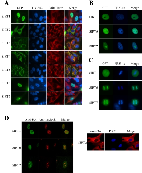
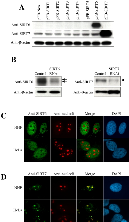
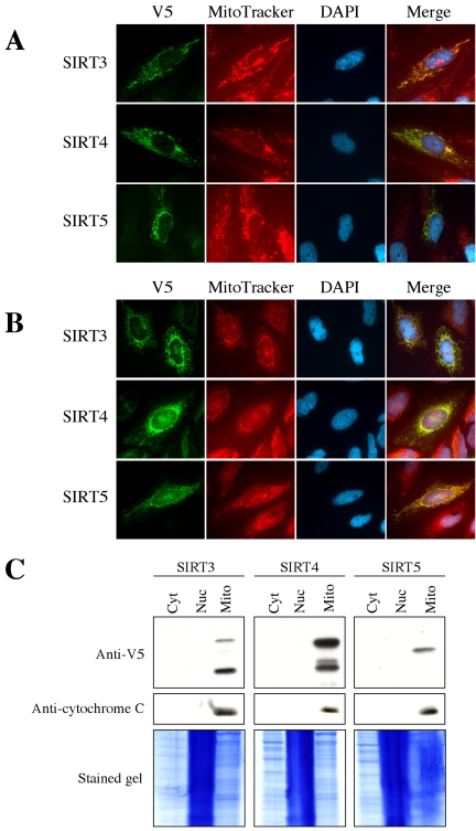
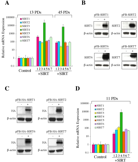
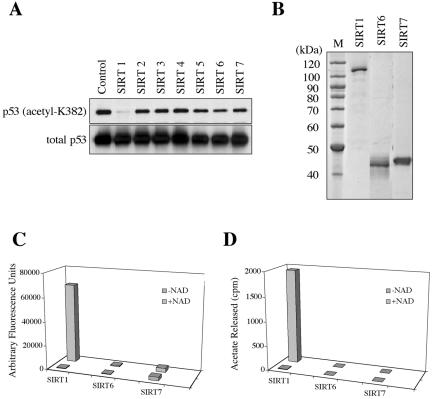
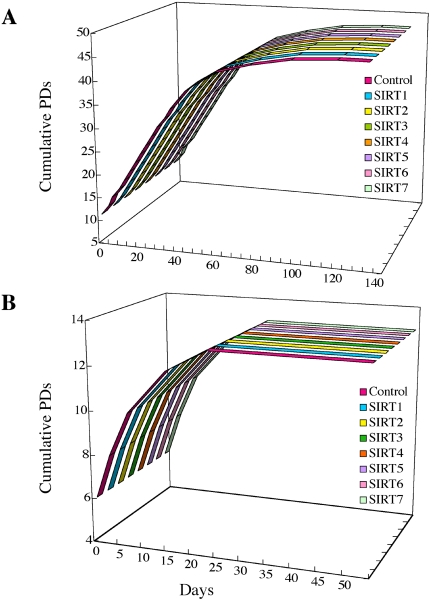
Sir2 is a NAD+-dependent protein deacetylase that extends lifespan in yeast and worms. This study examines seven human proteins homologous to Sir2 (SIRT1 through SIRT7) for cellular localization, expression profiles, protein deacetylation activity, and effects on human cell lifespan. We found that: 1) three nuclear SIRT proteins (SIRT1, SIRT6, and SIRT7) show different subnuclear localizations: SIRT6 and SIRT7 are associated with heterochromatic regions and nucleoli, respectively, where yeast Sir2 functions; 2) SIRT3, SIRT4, and SIRT5 are localized in mitochondria, an organelle that links aging and energy metabolism; 3) cellular p53 is a major in vivo substrate of SIRT1 deacetylase, but not the other six SIRT proteins; 4) SIRT1, but not the other two nuclear SIRT proteins, shows an in vitro deacetylase activity on histone H4 and p53 peptides; and 5) overexpression of any one of the seven SIRT proteins does not extend cellular replicative lifespan in normal human fibroblasts or prostate epithelial cells. This study supports the notion that multiple human SIRT proteins have evolutionarily conserved and nonconserved functions at different cellular locations and reveals that the lifespan of normal human cells, in contrast to that of lower eukaryotes, cannot be manipulated by increased expression of a single SIRT protein.


Figures

Figure 1.
Figure 1.
Cellular localization of human SIRT proteins. (A) Each SIRT-GFP fusion protein was transiently expressed in NHF normal human fibroblasts. Hoechst 33342 (H33342) and MitoFluor stainings are shown in parallel. The merged pictures of GFP, Hoechst 33342 and MitoFluor are in the most right panels. (B) Magnified nuclei of NHF cells expressing SIRT1-GFP, SIRT6-GFP, or SIRT7-GFP protein. (C) Mitotic NHF cells expressing SIRT1-GFP, SIRT6-GFP, or SIRT7-GFP protein. Condensed chromosomes are strongly stained with Hoechst 33342 in the middle panels. (D) NHF cells expressing HA-tagged SIRT1, SIRT2, SIRT6, or SIRT7 were immunostained with anti-HA antibody, in combination with anti-nucleoli antibody (for SIRT1, SIRT6, and SIRT7) or DAPI staining (for SIRT2).
Figure 2.
Figure 2.
Production of anti-SIRT6 and anti-SIRT7 antibodies and detection of endogenous SIRT6 and SIRT7 proteins. (A) NHF cells were infected with the retroviral vector driving each SIRT (pFB-SIRT1–7) or control vector (pFB-Neo) and used in Western blot with anti-SIRT6 and anti-SIRT7 rabbit polyclonal antibodies. β-actin was a loading control. (B) The endogenous SIRT6 (left) or SIRT7 (right) expression was knocked down by RNA interference (RNAi) in HeLa cells. The arrows indicate the specific signals, which were significantly weakened in the RNAi cells. The nature of two SIRT6 bands is currently unknown. (C and D) Indirect immunofluorescence staining of endogenous SIRT6 (C) and SIRT7 (D) proteins in NHF and HeLa cells. Costaining with anti-nucleoli antibody and DAPI staining are also shown.
Figure 3.
Figure 3.
Mitochondrial localization of SIRT3, SIRT4, and SIRT5. (A and B) The V5-His-tagged SIRT3, SIRT4, or SIRT5 was transiently expressed in NHF (A) and HeLa (B). The tagged proteins were detected by immunostaining with anti-V5 antibody. MitoTracker staining and DAPI staining are shown in parallel. (C) Cytoplasmic (Cyt), nuclear (Nuc), and mitochondrial (Mito) proteins were fractionated from 293T cells expressing V5-His-tagged SIRT3 (left), SIRT4 (middle), or SIRT5 (right), and analyzed in Western blot using anti-V5 antibody. Cytochrome C, a well known mitochondrial protein, was detected as a control. The gels stained with SimplyBlue SafeStain (Invitrogen) are also shown. Residual DNA in nuclear fractions gave the stronger staining. The presence of two SIRT3 bands is due to its N-terminal processing (Schwer et al., 2002). The exact nature of multiple SIRT4 bands is not known.
Figure 4.
Figure 4.
Establishment of normal human cells stably overexpressing each human SIRT. (A) NHF cells with the retroviral vector driving each SIRT (+SIRT1–7) at 13 PDs and 45 PDs were examined for mRNA expression of the respective SIRT gene by the real-time quantitative RT-PCR assays. NHF with the empty vector (Control) was also examined for the endogenous expression of each SIRT gene. The data are expressed as relative values to the endogenous expression levels in control cells (defined as 1 for each SIRT gene). (B) Western blot showed the overexpression of SIRT1, SIRT2, SIRT6, or SIRT7 protein in the respective retroviral vector-transduced NHF. The antibodies specific to each SIRT protein were used. (C) Western blot showed the overexpression of HA-SIRT1, HA-SIRT2, HA-SIRT6, or HA-SIRT7 protein in the respective retroviral vector-transduced NHF. Anti-HA antibody was used. (D) PrEC cells with the retroviral vector driving each SIRT (+SIRT1–7) or the empty vector (Control) were examined at 11 PDs as in A.
Figure 5.
Figure 5.
Protein deacetylation assays of human SIRT proteins. (A) Cellular p53 protein was immunoprecipitated from control and the SIRT-overexpressing NHF cells, followed by Western blot analysis using anti-acetyl-K382p53 antibody (top panel) and anti-p53 antibody (DO-1, bottom panel). The amounts of K382-acetylated p53 were normalized with those of total p53 protein detected by DO-1. In three independent experiments (a representative result shown here), the SIRT1-overexpressing cells, but not any others, showed a significant decrease in the acetylated p53. (B) Recombinant SIRT1, SIRT6, and SIRT7 proteins were purified from the baculovirus-infected insect cells. One microgram of proteins were analyzed on a SDS-polyacrylamide gel. (C) The purified SIRT1, SIRT6, and SIRT7 proteins were examined for an in vitro deacetylase activity on the synthetic p53 peptide (amino acid residues 379–382). The reactions with and without NAD+ were included. (D) The purified SIRT1, SIRT6, and SIRT7 proteins were examined for an in vitro deacetylase activity on the histone H4 peptide (amino acid residues 2–24) in the presence or absence of NAD+.
Figure 6.
Figure 6.
Replicative lifespan of NHF (A) and PrEC (B) overexpressing human SIRT proteins. The replicative histories of control and SIRT-overexpressing cells are shown as the growth curves, where the cumulative PDs are plotted to the days after retroviral transduction. All NHF-derived and PrEC-derived cells ceased to proliferate at 47 and at 13 PDs, respectively, with the characteristic features of cellular senescence such as enlarged and flat cell morphology and positive SA-β-gal staining.
Figure 7.
Figure 7.
Endogenous SIRT expression during replicative aging in normal human fibroblasts. (A) The real-time RT-PCR assays quantitated mRNA expression of each SIRT gene in NHF (at 23, 33, and 41 PDs) and WI-38 (at 34 PDs, 42 PDs, and 54 PDs). (B) The same set of cells as in (A) was examined in Western blot for SIRT1, SIRT6, and SIRT7 protein expression. β-actin was a loading control.
Figure 8.
Figure 8.
Expression profiles of SIRT genes in human organs. Twelve adult organs, fetal brain, and fetal liver were examined for the expression of each SIRT gene by the quantitative real-time RT-PCR assay. For each SIRT, the expression level in fetal brain was defined as 1, and the expression levels in the other organs were expressed as the relative values to fetal brain.

References

    1. Baptiste, N., and Prives, C. (2004). p53 in the cytoplasm: a question of overkill? Cell 116, 487–489. - PubMed
    1. Barbieri, M., Bonafe, M., Franceschi, C., and Paolisso, G. (2003). Insulin/IGF-I-signaling pathway: an evolutionarily conserved mechanism of longevity from yeast to humans. Am. J. Physiol. Endocrinol. Metab. 285, E1064–E1071. - PubMed
    1. Blander, G., and Guarente, L. (2004). The sir2 family of protein deacetylases. Annu. Rev. Biochem. 73, 417–435. - PubMed
    1. Bodnar, A. G., Ouellette, M., Frolkis, M., Holt, S. E., Chiu, C. P., Morin, G. B., Harley, C. B., Shay, J. W., Lichtsteiner, S., and Wright, W. E. (1998). Extension of life-span by introduction of telomerase into normal human cells. Science 279, 349–352. - PubMed
    1. Brooks, C. L., and Gu, W. (2003). Ubiquitination, phosphorylation and acetylation: the molecular basis for p53 regulation. Curr. Opin. Cell Biol. 15, 164–171. - PubMed

Publication types