. 2005 Nov;289(5):C1179-87.
doi: 10.1152/ajpcell.00062.2005. Epub 2005 Aug 3.

Role of TNF-{alpha} signaling in regeneration of cardiotoxin-injured muscle

Affiliations

Role of TNF-{alpha} signaling in regeneration of cardiotoxin-injured muscle

Shuen-Ei Chen et al. Am J Physiol Cell Physiol. 2005 Nov.

Abstract

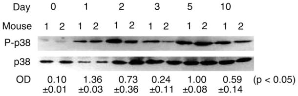
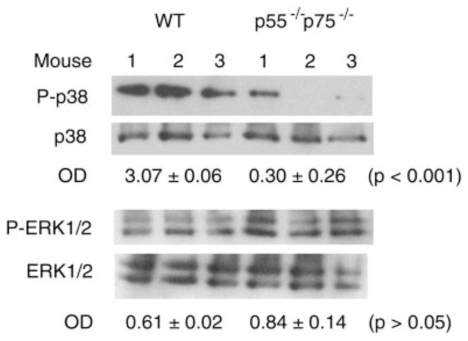
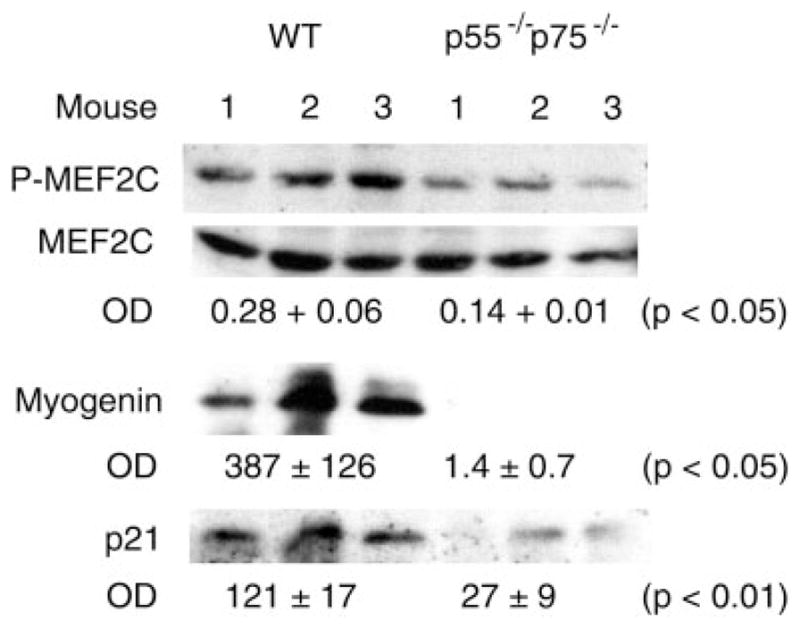
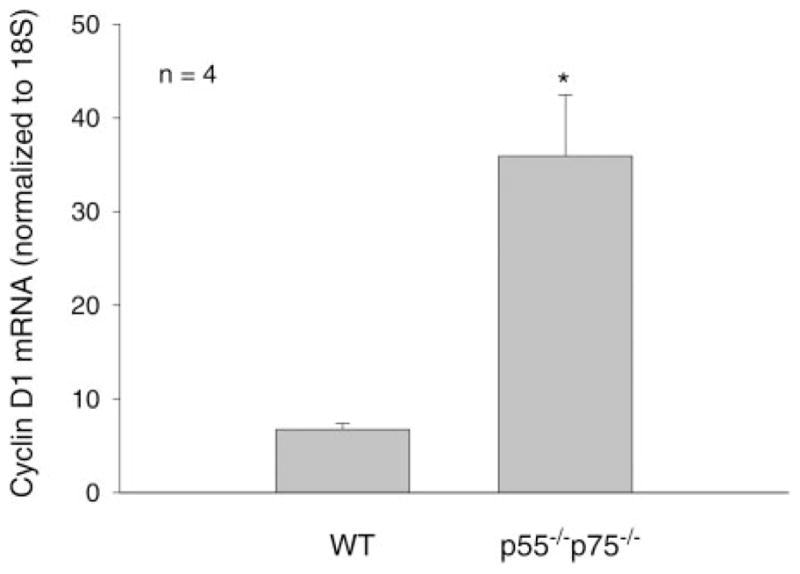
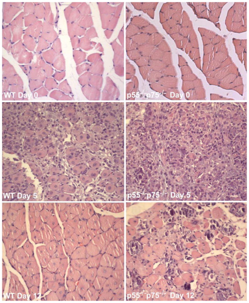
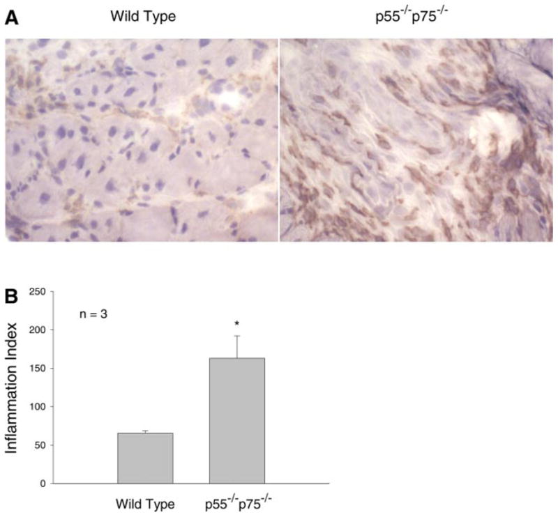
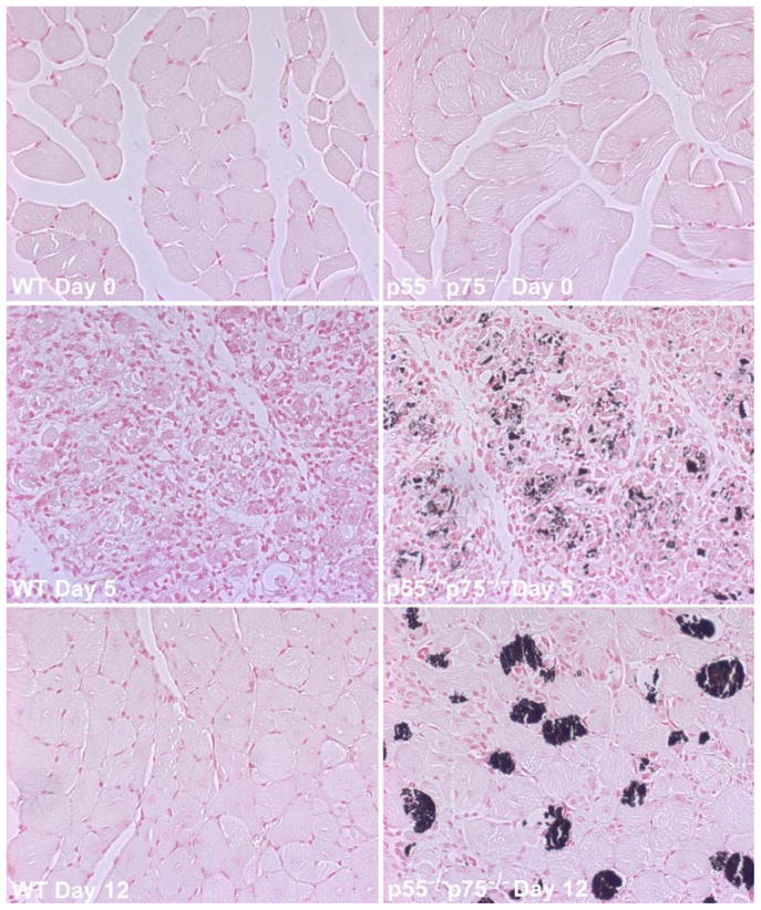
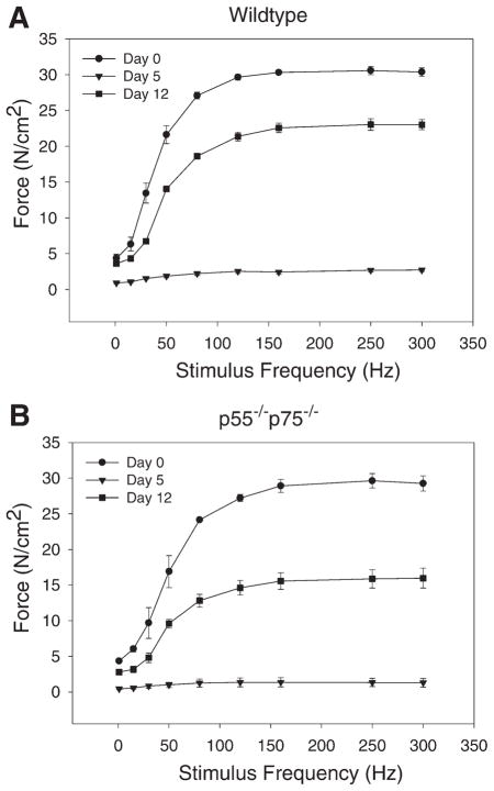
Recent data suggest a physiological role for the proinflammatory cytokine TNF-alpha in skeletal muscle regeneration. However, the underlying mechanism is not understood. In the present study, we analyzed TNF-alpha-activated signaling pathways involved in myogenesis in soleus muscle injured by cardiotoxin (CTX) in TNF-alpha receptor double-knockout mice (p55(-/-)p75(-/-)). We found that activation of p38MAPK, which is critical for myogenesis, was blocked in CTX-injured p55(-/-)p75(-/-) soleus on day 3 postinjury when myogenic differentiation was being initiated, while activation of ERK1/2 and JNK MAPK, as well as transcription factor NF-kappaB, was not reduced. Consequently, the phosphorylation of transcription factor myocyte enhancer factor-2C, which is catalyzed by p38 and crucial for the expression of muscle-specific genes, was blunted. Meanwhile, expression of p38-dependent differentiation marker myogenin and p21 were suppressed. In addition, expression of cyclin D1 was fivefold that in wild-type (WT) soleus. These results suggest that myogenic differentiation is blocked or delayed in the absence of TNF-alpha signaling. Histological studies revealed abnormalities in regenerating p55(-/-)p75(-/-) soleus. On day 5 postinjury, new myofiber formation was clearly observed in WT soleus but not in p55(-/-)p75(-/-) soleus. To the contrary, p55(-/-)p75(-/-) soleus displayed renewed inflammation and dystrophic calcification. On day 12 postinjury, the muscle architecture of WT soleus was largely restored. Yet, in p55(-/-)p75(-/-) soleus, multifocal areas of inflammation, myofiber death, and myofibers with smaller cross-sectional area were observed. Functional studies demonstrated an attenuated recovery of contractile force in injured p55(-/-)p75(-/-) soleus. These data suggest that TNF-alpha signaling plays a critical regulatory role in muscle regeneration.


Figures

Fig. 1
Fig. 1
Activation of p38 during regeneration of cardiotoxin (CTX)-injured mouse soleus muscle. Total protein was extracted from soleus collected from wild-type (WT) mice on the indicated days after being injured by direct injection of 100 μl of 10 μM CTX. Western blot analysis was performed using antibodies that are specific for phosphorylated p38 or pan-p38. Average optical density (OD) data are expressed as the ratio of phospho-p38 to p38 and were assessed using ANOVA.
Fig. 2
Fig. 2
Blockade of p38 activation in TNF-α receptor double-knockout mice (p55−/−p75−/−) soleus muscle. Total protein was extracted from soleus collected from WT and p55−/−p75−/− mice 3 days after the mice were injured using CTX. Western blot analysis was performed using antibodies that are specific for phosphorylated p38 or ERK1/2 and pan-p38 or pan-ERK1/2. OD data are expressed as the ratio of phosphorylated protein to total protein and were analyzed using Student’s t-test.
Fig. 3
Fig. 3
Blockade of p38-dependent myogenic signaling in regenerating p55−/−p75−/− soleus. Total protein or nuclear protein was extracted from soleus collected from WT and p55−/−p75−/− mice 3 days postinjury. Western blot analysis was performed using antibodies that are specific for phosphorylated myocyte enhancer factor (MEF)-2C and pan-MEF-2C to assess its activation in muscle nuclear extracts. Expression of myogenin and p21 was evaluated using Western blot analysis of muscle total protein extracts. OD data for MEF-2C activation are expressed as the ratio of phospho-MEF-2C to total MEF-2C, and OD data for myogenin and p21 expression are expressed as direct readings from the densitometer. Data were analyzed using Student’s t-test.
Fig. 4
Fig. 4
Deregulation of cyclin D1 expression in p55−/−p75−/− soleus. Total RNA was extracted from solei collected from WT and p55−/−p75−/− mice on day 3 after CTX injection. RT and real-time PCR were performed to evaluate cyclin D1 mRNA levels, which were normalized to 18S. The ratio of cyclin D1 expression to 18S is expressed in arbitrary units. Data are means ± SE derived from 5 mice in each group. *P < 0.01 (Student’s t-test).
Fig. 5
Fig. 5
Abnormal morphology in regenerating p55−/−p75−/− soleus muscle. Solei of adult WT and p55−/−p75−/− mice were injured by CTX injection and collected surgically on day 5 or day 12 after injection. Uninjured soleus was collected as control (day 0). Cross sections of formalin-fixed and paraffin-embedded soleus muscles were processed for hematoxylin and eosin staining. Representative sections of regenerating WT and p55−/−p75−/− soleus are shown.
Fig. 6
Fig. 6
Increased inflammation in regenerating p55−/−p75−/− soleus muscle. Cross sections of frozen soleus that were prepared from WT and p55−/−p75−/− mice on day 5 after CTX injection were stained with anti-Mac-1 (A). The average number of Mac-1-positive cells observed in a 0.25-mm2 area under a microscope was expressed as the inflammation index (B). Means ± SE derived from solei of 3 mice in each group are shown and were analyzed using Student’s t-test. *P < 0.05.
Fig. 7
Fig. 7
Calcification of regenerating p55−/−p75−/− soleus muscle. Solei of adult WT and p55−/−p75−/− mice were injured by direct injection of CTX and collected surgically on day 5 or day 12 after injection. Uninjured soleus was collected as control (day 0). Cross sections of formalin-fixed and paraffin-embedded soleus muscle were processed for Von Kassa staining, which stains solidified Ca2+ in black. Representative sections of regenerating WT and p55−/−p75−/− soleus are shown.
Fig. 8
Fig. 8
Compromised restoration of contractile force in injured p55−/−p75−/− soleus muscle. Soleus was excised from p55−/−p75−/− (B) and WT (A) mice before (day 0) or after CTX injection (days 5 and 12). Force developed by the muscle stimulated with 300-ms stimulus trains in Krebs-Ringer solution bath was recorded using an oscilloscope through a force transducer and normalized to muscle cross-sectional area (N/cm2). Data represent results from 3 or 4 mice (1 soleus per mouse) in each group. Data are means ± SE. The percentages of maximal force on day 12 compared with those on day 0 derived from WT and p55−/−p75−/− mice were compared using Student’s t-test.

References

    1. Aronson D, Wojtaszewski JFP, Thorell A, Nygren J, Zangen D, Richter EA, Ljungqvist O, Fielding RA, Goodyear LJ. Extracellular-regulated protein kinase cascades are activated in response to injury in human skeletal muscle. Am J Physiol Cell Physiol. 1998;275:C555–C561. - PubMed
    1. Cabane C, Englaro W, Yeow K, Ragno M, Dérijard B. Regulation of C2C12 myogenic terminal differentiation by MKK3/p38α pathway. Am J Physiol Cell Physiol. 2003;284:C658–C666. - PubMed
    1. Cantini M, Carraro U. Macrophage-released factor stimulates selectively myogenic cells in primary muscle culture. J Neuropathol Exp Neurol. 1995;54:121–128. - PubMed
    1. Cantini M, Massimino ML, Rapizzi E, Rossini K, Catani C, Dalla Libera L, Carraro U. Human satellite cell proliferation in vitro is regulated by autocrine secretion of IL-6 stimulated by a soluble factor(s) released by activated monocytes. Biochem Biophys Res Commun. 1995;216:49–53. - PubMed
    1. Chazaud B, Sonnet C, Lafuste P, Bassez G, Rimaniol AC, Poron F, Authier FJ, Dreyfus PA, Gherardi RK. Satellite cells attract monocytes and use macrophages as a support to escape apoptosis and enhance muscle growth. J Cell Biol. 2003;163:1133–1143. - PMC - PubMed

Publication types

MeSH terms

Substances

LinkOut - more resources