

Roles of PSF protein and VL30 RNA in reversible gene regulation
- PMID: 16079199
- PMCID: PMC1189330
- DOI: 10.1073/pnas.0505179102
Roles of PSF protein and VL30 RNA in reversible gene regulation
Erratum in
- Proc Natl Acad Sci U S A. 2005 Nov 15;102(46):16905
Abstract
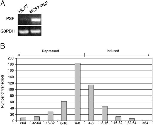
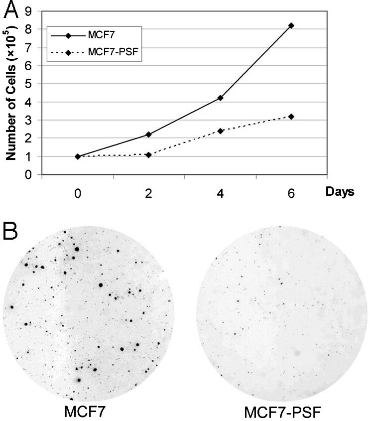
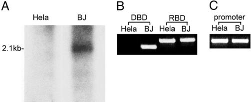
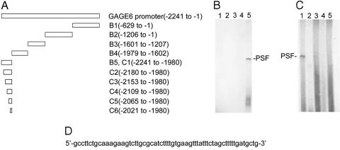
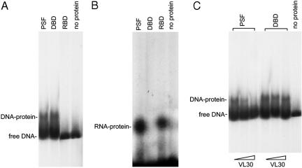
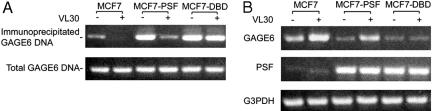
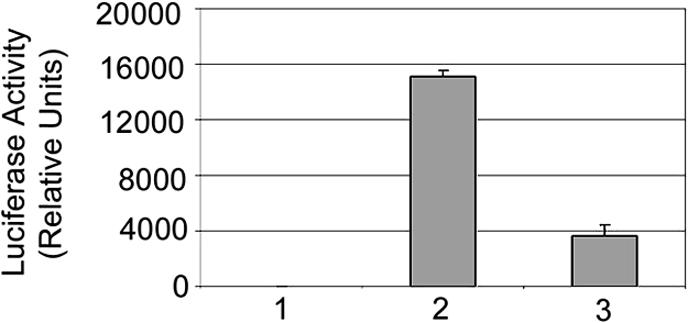
The mammalian protein PSF contains a DNA-binding domain (DBD) that coordinately represses multiple oncogenic genes in human cell lines, indicating a role for PSF as a human tumor-suppressor protein. PSF also contains two RNA-binding domains (RBD) that form a complex with a noncoding VL30 retroelement RNA, releasing PSF from a gene and reversing repression. Thus, the DBD and RBD in PSF are linked by a mechanism of reversible gene regulation involving a noncoding RNA. This mechanism also could apply to other regulatory proteins that contain both DBD and RBD. The mouse genome has multiple copies of VL30 retroelements that are developmentally regulated, and mouse cells contain VL30 RNAs that have normal and pathological roles in gene regulation. Human chromosome 11 has a VL30 retroelement, and a VL30 EST was identified in human blastocyst cells, indicating that the PSF-VL30 RNA regulatory mechanism also could function in human cells.
Figures

Comment in
-
Normal and pathological functions of mammalian retroelements.Proc Natl Acad Sci U S A. 2005 Aug 30;102(35):12292-3. doi: 10.1073/pnas.0505866102. Epub 2005 Aug 23. Proc Natl Acad Sci U S A. 2005. PMID: 16118280 Free PMC article. Review. No abstract available.
Similar articles
-
Regulation of proto-oncogene transcription, cell proliferation, and tumorigenesis in mice by PSF protein and a VL30 noncoding RNA.Proc Natl Acad Sci U S A. 2009 Sep 29;106(39):16794-8. doi: 10.1073/pnas.0909022106. Epub 2009 Sep 11. Proc Natl Acad Sci U S A. 2009. PMID: 19805375 Free PMC article.
-
Binding of mouse VL30 retrotransposon RNA to PSF protein induces genes repressed by PSF: effects on steroidogenesis and oncogenesis.Proc Natl Acad Sci U S A. 2004 Jan 13;101(2):621-6. doi: 10.1073/pnas.0307794100. Epub 2004 Jan 2. Proc Natl Acad Sci U S A. 2004. PMID: 14704271 Free PMC article.
-
From a retrovirus infection of mice to a long noncoding RNA that induces proto-oncogene transcription and oncogenesis via an epigenetic transcription switch.Signal Transduct Target Ther. 2016 May 13;1:16007. doi: 10.1038/sigtrans.2016.7. eCollection 2016. Signal Transduct Target Ther. 2016. PMID: 29263895 Free PMC article. Review.
-
Role of human noncoding RNAs in the control of tumorigenesis.Proc Natl Acad Sci U S A. 2009 Aug 4;106(31):12956-61. doi: 10.1073/pnas.0906005106. Epub 2009 Jul 22. Proc Natl Acad Sci U S A. 2009. PMID: 19625619 Free PMC article.
-
PSF: nuclear busy-body or nuclear facilitator?Wiley Interdiscip Rev RNA. 2015 Jul-Aug;6(4):351-67. doi: 10.1002/wrna.1280. Epub 2015 Apr 1. Wiley Interdiscip Rev RNA. 2015. PMID: 25832716 Free PMC article. Review.
Cited by
-
Rab23 activities and human cancer-emerging connections and mechanisms.Tumour Biol. 2016 Oct;37(10):12959-12967. doi: 10.1007/s13277-016-5207-7. Epub 2016 Jul 23. Tumour Biol. 2016. PMID: 27449041 Review.
-
Interaction of host cellular proteins with components of the hepatitis delta virus.Viruses. 2010 Jan;2(1):189-212. doi: 10.3390/v2010189. Epub 2010 Jan 18. Viruses. 2010. PMID: 21994607 Free PMC article.
-
VL30 retrotransposition is associated with induced EMT, CSC generation and tumorigenesis in HC11 mouse mammary stem‑like epithelial cells.Oncol Rep. 2020 Jul;44(1):126-138. doi: 10.3892/or.2020.7596. Epub 2020 Apr 28. Oncol Rep. 2020. PMID: 32377731 Free PMC article.
-
The nucleic acid binding protein SFPQ represses EBV lytic reactivation by promoting histone H1 expression.Nat Commun. 2024 May 16;15(1):4156. doi: 10.1038/s41467-024-48333-x. Nat Commun. 2024. PMID: 38755141 Free PMC article.
-
The DBHS proteins SFPQ, NONO and PSPC1: a multipurpose molecular scaffold.Nucleic Acids Res. 2016 May 19;44(9):3989-4004. doi: 10.1093/nar/gkw271. Epub 2016 Apr 15. Nucleic Acids Res. 2016. PMID: 27084935 Free PMC article.
References
-
- Patton, J. G., Porro, E. B., Galceran, J., Tempst, P. & Nadalginard, B. (1993) Genes Dev. 7, 393-406. - PubMed
-
- Urban, R. J., Bodenburg, Y., Kurosky, A., Wood, T. G. & Gasic, S. (2000) Mol. Endocrinol. 14, 774-782. - PubMed
-
- Urban, R. J., Bodenburg, Y. H. & Wood, T. G. (2002) Am. J. Physiol. 283, E423-E427. - PubMed
Publication types
MeSH terms
Substances
LinkOut - more resources
Full Text Sources
Research Materials
