. 2005 Oct;16(10):617-24.
doi: 10.1016/j.jnutbio.2005.03.006.

HepG2 cells develop signs of riboflavin deficiency within 4 days of culture in riboflavin-deficient medium

Affiliations

HepG2 cells develop signs of riboflavin deficiency within 4 days of culture in riboflavin-deficient medium

Ricarda Werner et al. J Nutr Biochem. 2005 Oct.

Abstract

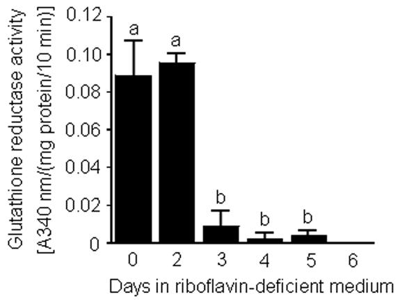
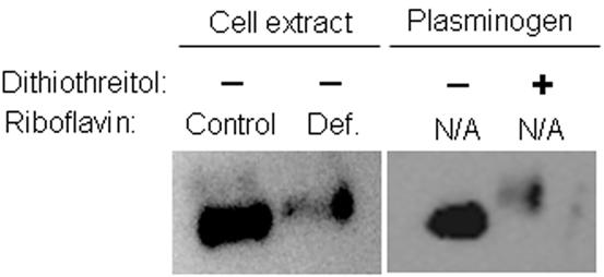
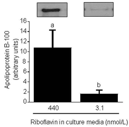
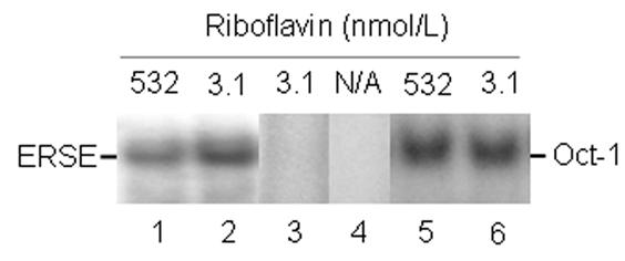
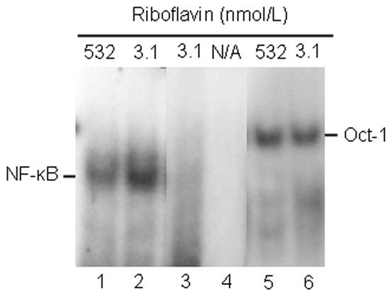
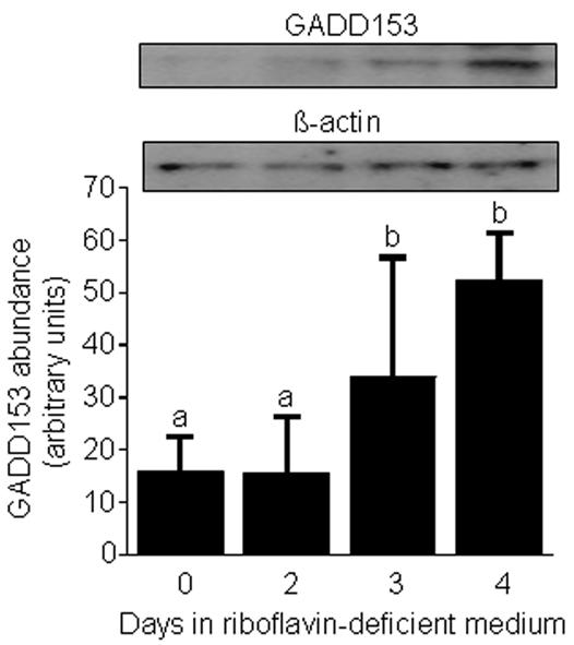
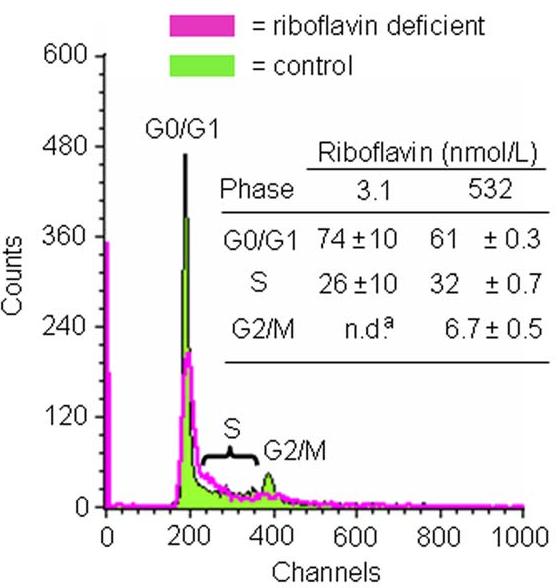
Flavin mononucleotide (FMN) and flavin adenine dinucleotide (FAD) are essential coenzymes in redox reactions. For example, FAD is a coenzyme for both glutathione reductase and enzymes that mediate the oxidative folding of secretory proteins. Here we investigated short-term effects of moderately riboflavin-deficient culture medium on flavin-related responses in HepG2 hepatocarcinoma cells. Cells were cultured in riboflavin-deficient (3.1 nmol/l) medium for up to 6 days; controls were cultured in riboflavin-sufficient (532 nmol/l) medium. The activity of glutathione reductase decreased by 98% within 4 days of riboflavin-deficient culture. Transport rates of riboflavin increased in response to riboflavin depletion, whereas expression of enzymes mediating flavocoenzyme synthesis (flavokinase and FAD synthetase) decreased in response to depletion. The oxidative folding and synthesis of plasminogen and apolipoprotein B-100 was impaired within 4 days of culture in riboflavin-deficient medium; this is consistent with impaired processing of secretory proteins in riboflavin-deficient cells. Riboflavin depletion was associated with increased DNA-binding activities of transcription factors with affinity for endoplasmic reticulum stress elements and nuclear factor kappaB (NF-kappaB) consensus elements, suggesting cell stress. Moreover, the abundance of the stress-induced protein GADD153 was greater in riboflavin-deficient cells compared with controls. Riboflavin deficiency was associated with decreased rates of cell proliferation caused by arrest in G1 phase of the cell cycle. These studies are consistent with the hypothesis that HepG2 cells have a great demand for riboflavin and that cell stress develops rapidly if riboflavin supply is marginally low.


Figures

Fig. 1.
Fig. 1.
Time course of glutathione reductase activity in HepG2 cells in response to transfer "from riboflavin-sufficient medium (532 nmol/L, day 0) to riboflavin-deficient medium (3.1 nmol/L). a,bBars not sharing the same letter are significantly different (P < 0.05; n = 4). Means ± SD are shown.
Fig. 2
Fig. 2
The abundance of mRNA coding for flavokinase and FAD synthetase decreases in response to riboflavin deficiency in HepG2 cells. Cells were cultured in riboflavin-deficient (3.1 nmol/L) and riboflavin-sufficient (532 nmol/L) media for 4 d. mRNA abundance was quantified using RT-PCR. Data are expressed in units of percent mRNA abundance, using cells cultured in medium containing 532 nmol/L riboflavin as the reference. a,bBars not sharing the same letter are significantly different (P < 0.05; n = 4). Means ± SD are shown.
Fig. 3
Fig. 3
The abundance of free sulfhydryl groups is greater in proteins from riboflavin-deficient HepG2 cells (“Def.” = 3.1 nmol/L riboflavin) compared with riboflavin-sufficient controls (“Control” = 532 nmol/L riboflavin). Cells were cultured in riboflavin-defined media for 4 d. Sulfhydryl groups in proteins from cell extracts were derivatized with polyethylene oxide-iodoacetyl biotin; plasminogen was visualized by Western blot analysis using an antibody to human plasminogen. Commercial, chemically pure plasminogen (with and without pretreatment with dithiothreitol) was used as a control. N/A = not applicable.
Fig. 4
Fig. 4
Riboflavin deficiency decreases the abundance of apolipoprotein B-100 in HepG2 cells. Cells were cultured in riboflavin-deficient (3.1 nmol/L) and riboflavin-sufficient (532 nmol/L) media for 4 d. Apolipoprotein B-100 was quantified by Western blot analysis and gel densitometry. Inserts depict representative Western blot images. a,bBars not sharing the same letter are significantly different (P < 0.05; n = 4). Means ± SD are shown.
Fig. 5
Fig. 5
Riboflavin deficiency enhances binding of transcription factors to endoplasmic reticulum stress elements in HepG2 cells. Cells were cultured in riboflavin-deficient (3.1 nmol/L) and riboflavin-sufficient (532 nmol/L) media for 4 d. Binding to endoplasmic reticulum stress elements was visualized using EMSA; binding to an Oct-1 site was used as a control. N/A = not applicable. Lanes 1 and 2 = binding to an endoplasmic reticulum stress element by nuclear extracts from control cells and riboflavin-deficient cells, respectively; lane 3 = 100-fold molar excess of unlabeled endoplasmic reticulum stress element; lane 4 = endoplasmic reticulum stress element in the absence of nuclear extract; lanes 5 and 6 = binding to a consensus binding sequence for Oct-1 by nuclear extracts from control cells and riboflavin-deficient cells, respectively.
Fig. 6
Fig. 6
The DNA-binding activity of NF-κB increases in response to riboflavin deficiency in HepG2 cells. Cells were cultured in riboflavin-deficient (3.1 nmol/L) and riboflavin-sufficient (532 nmol/L) media for 4 d. Binding of NF-κB to response elements was visualized using EMSA; binding to an Oct-1 site was used as a control. N/A = not applicable. Lanes 1 and 2 = binding to a consensus sequence for NF-κB by nuclear extracts from control cells and riboflavin-deficient cells, respectively; lane 3 = 100-fold molar excess of unlabeled consensus binding site for NF-κB; lane 4 = consensus binding site for NF-κB in the absence of nuclear extract; lanes 5 and 6 = binding to a consensus binding sequence for Oct-1 by nuclear extracts from control cells and riboflavin-deficient cells, respectively.
Fig. 7
Fig. 7
The abundance of GADD153 increases in response to riboflavin deficiency in HepG2 cells. Cells were transferred from riboflavin-sufficient medium (532 nmol/L, day 0) to riboflavin-deficient medium (3.1 nmol/L). At timed intervals, GADD153 and β-actin (control) were quantified by Western blot analysis and gel densitometry. Inserts depict representative Western blot images. a,bBars not sharing the same letter are significantly different (P < 0.05; n = 4). Means ± SD are shown.
Fig. 8
Fig. 8
Riboflavin deficiency causes G0/G1 phase arrest in HepG2 cells. Cells were cultured in media containing 3.1 and 532 nmol/L riboflavin for 4 d. Cell cycle phase distribution was quantified by flow cytometry. Representative flow cytometry charts are depicted. The insert provides a table with percent cell cycle phase distributions. n.d. = not detectable. aSignificantly different from riboflavin-sufficient controls (P < 0.01; n = 3).

References

    1. Rivlin RS. Riboflavin. In: Present Knowledge in Nutrition. In: Bowman BA, Russell RM, editors. ILSI Press; Washington, DC: 2001.
    1. Tu BP, Ho-Schleyer SC, Travers KL, Weissman JS. Biochemical basis of oxidative protein folding in the endoplasmic reticulum. Science. 2000;290:1571–1574. - PubMed
    1. Thorpe C, Hoober K, Raje S, Glynn N, Burnside J, Turi G, Coppock D. Sulfhydryl oxidases: emerging catalysts of protein disulfide bond formation in eukaryotes. Arch. Bioch. Biophys. 2002;405:1–12. - PubMed
    1. Camporeale G, Zempleni J. Oxidative folding of interleukin-2 is impaired in flavin-deficient Jurkat cells, causing intracellular accumulation of interleukin-2 and increased expression of stress response genes. J. Nutr. 2003;133:668–672. - PubMed
    1. Manthey KC, Chew YC, Zempleni J. Riboflavin deficiency impairs oxidative folding and secretion of apolipoprotein B-100 in HepG2 cells, triggering stress-response systems. J. Nutr. 2005 in press. - PMC - PubMed

Publication types

MeSH terms