. 2005 Aug 16;102(33):11716-21.
doi: 10.1073/pnas.0501162102. Epub 2005 Aug 4.

Genetic reprogramming of tumor cells by zinc finger transcription factors

Affiliations

Genetic reprogramming of tumor cells by zinc finger transcription factors

Pilar Blancafort et al. Proc Natl Acad Sci U S A. .

Abstract

Cancer arises by the accumulation of genetic alterations in DNA leading to aberrant gene transcription. Expression-profiling studies have correlated genomewide expression signatures with malignancy. However, functional analysis elucidating the contribution and synergy of genes in specific cancer cell phenotypes remains a formidable obstacle. Herein, we describe an alternative genetic approach for identification of genes involved in tumor progression by using a library of zinc finger artificial transcription factors (ATFs) and functional screening of tumor cells as a source of genetic plasticity and clonal selection. We isolated a six-zinc finger transcriptional activator (TF 20-VP, TF 20 containing the VP64 activator domain) that acts to reprogram a drug-sensitive, poorly invasive, and nonmetastatic cell line into a cell line with a drug-resistant, highly invasive, and metastatic phenotype. Differential expression profiles of cells expressing TF 20-VP followed by functional studies, both in vitro and in animal models, revealed that invasion and metastasis requires co-regulation of multiple target genes. Significantly, the E48 antigen, associated with poor metastasis-free survival in head and neck cancer, was identified as one specific target of TF 20-VP. We have shown phenotypic modulation of tumor cell behavior by E48 expression, including enhanced cell migration in vitro and tumor cell dissemination in vivo. This study demonstrates the use of ATFs to identify the group of genes that cooperate during tumor progression. By co-regulating multiple targets, ATFs can be used as master genetic switches to reprogram and modulate complex neoplastic phenotypes.


Figures

Fig. 1.
Fig. 1.
TF 20 is able to induce complex phenotypes in HeLa cells. (A) Illustration of TF-mediated reprogramming of cancer cells. TF 20-VP induces morphological transformations and cytoskeleton remodeling in HeLa cells. (B) Aspect of a colony of control cells expressing no ZFs. (Magnification: ×100.) TF 20-SKD-transduced cells formed the same WT compact colonies (data not shown). (C and F) Control cells (C) and TF 20-VP-transduced cells (F) were stained for F-actin (Texas red-phalloidin, red) and nucleus (DAPI, blue). (Magnification: ×600.) (D and E) Morphological transformations of HeLa cells transduced with a retrovirus expressing TF 20-VP, showing cells migrating out of the colony. (Magnification: ×100.) (G) TF 20-VP enhances the migration of HeLa cells on a laminin migration assay.
Fig. 2.
Fig. 2.
TF 20-VP enhances cell invasion and metastasis. (A) Invasion assays with control and TF-transduced cells were performed in vitro by using Matrigel chambers. (B) TF 20-VP increased the number of lung micrometastases in a NOD SCID mouse model of spontaneous metastasis. Groups represent HeLa cells expressing TF 20 VP64 activator domain (TF 20-VP), the same protein but linked to a repressor domain (TF 20-SKD), and cells expressing no ZF domains (control). P values between TF 20-VP and control and between TF 20-VP and TF 20-SKD were <0.05; P values between TF 20-SKD and control were >0.4. (Magnifications: ×200.)
Fig. 3.
Fig. 3.
TF 20-VP regulates the endogenous E48 gene, a marker of disseminated tumor cells of squamous cell carcinomas of the head and neck. (A) HeLa cells were analyzed for E48 expression 72 h posttransduction. (B) HeLa cells recovered from mouse primary tumors 35 days postinjection. Flow cytometry analyses were performed with a mAb that detects human E48. E48 is up-regulated in HeLa cells transduced with a TF 20-VP retrovirus (red); HeLa cells expressing TF 20-SKD (light blue); control cells expressing no ZFs (green); untransduced cells (filled blue). (C) Immunofluorescence analysis of E48 expression in HeLa cells expressing TF 20-VP, control cells expressing no ZFs (control), and cells overexpressing an E48 cDNA (E48). Expression of E48 was induced in the cell–cell junctions (arrow). (Magnifications: ×400.) (D) Ectopic expression of E48 induces cell migration in Laminin-coated transwells. HeLa cells were transduced with EGFP, TF 20-VP, and E48 cDNA (E48). Untransduced (HeLa cells) were also evaluated. (E) HeLa cells transduced with TF 20-VP, TF 20-SKD, and E48 retroviruses were injected i.v. in chicken embryos; disseminated tumor cells were detected in distal organs (lung and lower chorioallantoic membrane, CAM) by real-time alu-PCR as described (27). HeLa cells expressing both TF 20-VP and E48 cDNA enhance 10- to 20-fold and 5-fold, respectively, the number of experimental metastasis in a chicken embryo model of organ colonization.
Fig. 4.
Fig. 4.
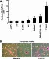
In vitro induction of cell invasion requires coexpression of multiple targets. (A) HeLa cells were transiently transfected with individual cDNAs encoding E48, AGT, IL-13Rα1, and TF 20-VP. Transfected cells were loaded into Matrigel chambers, and invading cells were fixed and counted. Values represent averages of two wells, and experiments were done in triplicate. (B) E48 and AGT coexpression in HeLa cells suffices to recapitulate the cell morphology changes mediated by TF 20-VP. (Magnification: ×400.)

References

    1. Ramaswamy, S., Ross, K. N., Lander, E. S. & Golub, T. R. (2003) Nat. Genet. 33, 49–87. - PubMed
    1. Rhodes, D. R., Yu, J., Shanker, K., Deshpande, N., Varambally, R., Ghosh, D., Barrette. T., Pandey, A. & Chinnaiyan, A. M. (2004) Proc. Natl. Acad. Sci. USA 101, 9309–9314. - PMC - PubMed
    1. Segal, E., Friedman, N. & Regev, A. (2004) Nat. Genet. 36, 1090–1098. - PubMed
    1. Beneklli, M., Baer, M. R., Baumann, H. & Wetzier, M. (2003) Blood 101, 2940–2951. - PubMed
    1. Kang, Y. & Massague, J. E. (2004) Cell 118, 277–279. - PubMed

Publication types

Substances