. 2005 Nov-Dec;23(10):1560-70.
doi: 10.1634/stemcells.2004-0284. Epub 2005 Aug 4.

Human umbilical cord blood progenitors: the potential of these hematopoietic cells to become neural

Affiliations

Human umbilical cord blood progenitors: the potential of these hematopoietic cells to become neural

Ning Chen et al. Stem Cells. 2005 Nov-Dec.

Abstract

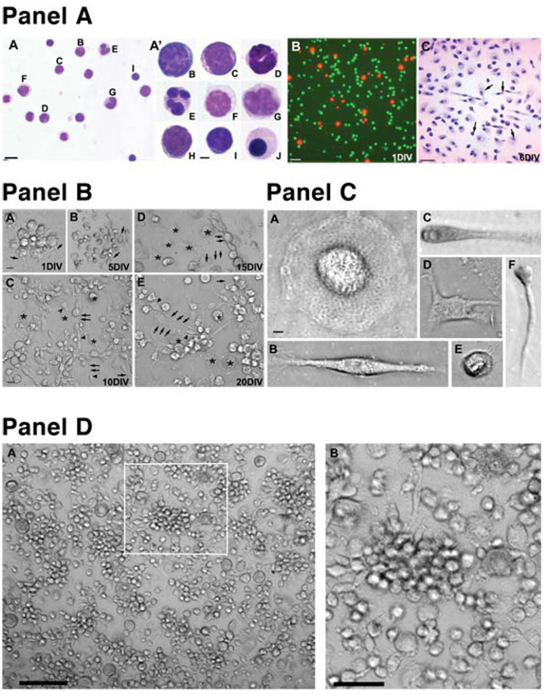
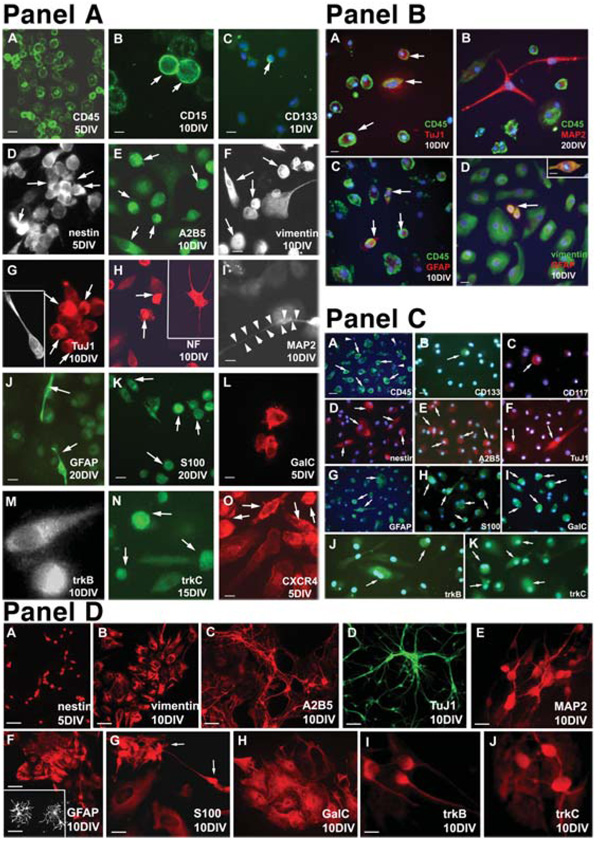
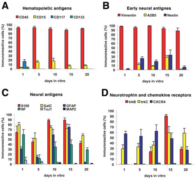
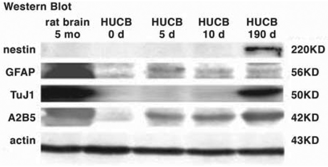
The mononuclear fraction from human umbilical cord blood (HUCB) contains a significant number of stem/progenitor cells that in theory could be come any cell in the body, including neurons. Taking into consideration that transdifferentiation would be a very rare event and also knowing that overlapping genetic programs for hematopoiesis and neuropoiesis exist, we undertook a characterization of the HUCB mononuclear fraction, including analysis of cellular subpopulations and their morphology, cell viability, proliferation, and expression of neural and hematopoietic antigens. Two cell populations were apparent-adherent and floating fractions. The adherent fraction was mainly lymphocytes (~53%) expressing hematopoietic antigens. Upon replate, the floating population had many cells that expressed stem cell antigens. More of the cells in this subfraction expressed neural proteins. Neurotrophin receptors trkB and trkC were present in both cell fractions, although expression was higher in the floating fraction. Our initial characterization suggests that a subpopulation of cells exists within the HUCB mononuclear fraction that seems to have the potential to become neural cells, which could then be used in the development of cell-based therapies for brain injuries and diseases.


Figures

Figure 1
Figure 1
The Wright-Giemsa staining and morphology of a mononuclear HUCB fraction before and after culturing. (Panel A): Part A, Giemsa stained smear of HUCB cells. Scale bar = 10 µm. Part A' shows high magnification of individual cell types depicted on the previous image: (B) myeloblast, (C) promyelocyte, (D) band, (E) neutrophil, (F) promonocyte, (G) monocyte, (H) lymphoblast, (I) small lymphocyte, and (J) orthochromic normoblast. Scale bar = 2 μm. Part B shows viability of cultured mononuclear HUCB cells; living cultures were stained with fluorescein diacetate/ propidium iodide to detect healthy (green) and dead (red) cells. Part C shows bright-field photomicrograph of Giemsastained cultured HUCB cells (6 DIV) demonstrating heterogeneous morphologies (arrows). Scale bar = 20 µm. (Panel B): Morphology of the adherent population of mononuclear HUCB cells cultured for 20 days. (A): One day after plating, cells (arrowheads) formed small clusters from which a few cells sent out tiny processes (arrows). During the next culture period (5–10 days), the clusters became loosely packed and many cells extended longer processes interconnecting cell groups. (B, C): Besides small round cells either with or without processes (arrows), numerous larger round cells (asterisks) were found. These cells were always tightly adhered to the bottom of the culture well. (D, E): In the next two time periods (15 and 20 DIV), small cells (arrowheads) with longer processes were interspersed either with individual or groups of round flat cells (asterisks). Scale bar = (A) 10 µm and (B–E) 20 µm. (Panel C): Different cell types were observed in adherent cultures: (A) large egg-shaped cells; (B) bipolar and (D) multipolar cells; (E) small round cells with multiple hair-like spines; and small cells with (C) thicker or (F) thinner processes. Scale bar = 2 µm. (Panel D): The morphology of cultured HUCB cells prepared from floating fractions. Morphologically, the cells were similar to the adherent population, with a striking absence of big round egg-like cells. (A): Numerous clusters (arrow) of uniform round cells with occasional processes were observed. (B): Higher magnification of the boxed area from (A). Scale bar = (A) 40 µm and (B) 20 µm. Abbreviations: DIV, days in vitro; HUCB, human umbilical cord blood.
Figure 2
Figure 2
Fluorescent images. (Panel A): Immunocytochemical characterization of (A–C) adherent mononuclear HUCB cells showing the expression of hematopoietic CD antigens. (A): Almost every cell revealed surface CD45 (green) antigen at 1, 5, 10, 15, and 20 days. (B): Only a few cells were positive for the antibody against CD15 (green, arrows) throughout the entire course of the study. (C): The presence of immature progenitors expressing CD133 (green, arrow) was recorded only in short-term (1 DIV) cultures. DAPI counterstaining was used for visualization of the entire cell population. Scale bar = (A, B) 20 µm and (C) 10 µm. (D–F): Several early neural markers were expressed in HUCB cells. Cells positive for (D) nestin, (E) A2B5, and (F) vimentin were observed throughout the entire culturing period. Arrows in (D–F) point to immunoreactive cells. (G–I): HUCB cells expressed early and mature neuronal antigens. Ten days after culturing, only a few cells expressed TuJ1 (G, arrows) and MAP2 (I). The arrow points to the MAP2-positive soma, whereas arrow-heads delineate the long process. (H): At the same time, many cells were immunoreactive for NF68KD (arrows). Scale bar = (D–I) 20 µm. (J–L): Cultured HUCB cells express glial antigens. (J): Numerous cells were positive for GFAP. The arrows point to two cells with bipolar morphology. In sister cultures, the positivity for other glial markers such as S100 (K, arrows) and GalC (L) was detected. Scale bar = (J, K) 20 µm and (L) 10 µm. (M–O): Expression of neurotrophin and chemokine receptors. (M): Positivity for trkB was found on day 5 only, whereas trkC (N, arrows) was present throughout the whole culture period. (O): CXCR4 was present in numerous cells in all studied intervals (arrows). Scale bar = (M–O) 10 µm. (Panel B): Coexpression of hematopoietic and neural antigens in cultured HUCB cells. (A): Several CD45-positive (green) cells coexpressing TuJ1 (red). Hematopoietic CD markers are localized on the cell surface (arrows) of many HUCB cells, whereas weak cytoplasmic TuJ1 expression was found occasionally. Nuclear DAPI labeling (blue) confirms that not every HUCB cell expresses CD antigens. (B): For longer cultures, a cell with distinct processes positive for mature neuronal marker MAP2 (red) and negative for CD45. On the other hand, cells with round morphology were CD45 immunoreactive (green). (C): Coexpression of GFAP (red) and CD45 (green) was also found (arrows). (D): HUCB cells were immunopositive for vimentin (green), and some also expressed GFAP (red). The arrow indicates vimentin/GFAP-postive cells. Inset shows a double-labeled vimentin/GFAP-positive (orange) cell at a higher magnification. Scale bar = (A–C, inset in D) 10 µm and (D) 20 µm. (Panel C): Fluorescent images of replated floating HUCB cells. (A–C): Hematopoietic markers, including (A) CD45, (B) CD133, and (C) CD117. Arrowheads point to cells negative for the specific marker, whereas arrows indicate cells expressing one of the CD antigens. (D, E): Expression of (D) nestin and (E) A2B5 (arrows) was higher in the replated floating fraction than in the adhered HUCB cells. (F–I): Cells positive for (F) TuJ1, (G) GFAP, (H) S100, and (I) GalC. Morphologically, this population of cells rarely acquired the classical appearance of neural derivatives. (J, K): Expression of neurotrophin receptors (J) trkB and (K) trkC was also detected. Arrows in (F–K) point to positively labeled cells. Blue DAPI counterstaining was used for clear identification of all cultured cells. Scale bar = (A, D) 20 µm and (B, C, E–K) 40 µm. (Panel D): Expression of neural antigens in E14–E18 primary cultures. (A–C): Cells isolated from the embryonic striatum were immunopositive for (A) nestin and (B) vimentin, and cultures prepared from the embryonic olfactory bulb revealed positivity for (C) A2B5. Scale bar = (A) 10 µm and (B, C) 40 µm. TuJ1 (D) and MAP2 (E) were found in cortical cultures after 10 days. Cells expressing these two neuronal antigens revealed classical neuronal morphology. (F): Small neuronal bodies and long, branching processes differed from flat, larger cells expressing GFAP. Inset shows the detailed morphology of GFAP-positive cells taken from E18 striatal culture. Scale bar = (D) 40 µm, (E) 10 µm, and 20 µm. Scale bar in inset = 10 µm. (G, H): Cortex-derived cells also expressed two other glial antigens. Arrows in (G) point to S100-positive cells. Scale bar = 20 µm. (I, J): Cortical cells also reveal positivity for neurotrophin receptors. Scale bar = 10 µm. Abbreviations: DAPI, 4,6-diamidino-2-phenylindole; DIV, days in vitro; HUCB, human umbilical cord blood.
Figure 3
Figure 3
Antigen expression in adherent HUCB cells cultured in serum from 1–20 days in vitro. (A): Expression of early hematopoietic and stem cell antigens. (B): Expression of early neural/stem cell antigens. (C): Expression of antigens indicative of immature and mature neurons, astrocytes, and oligodendrocytes. (D): The adherent HUCB cells also expressed both neurotrophin receptors and the receptor for the chemokine stromal-derived factor 1, CXCR4. Abbreviation: HUCB, human umbilical cord blood.
Figure 4
Figure 4
Western blot analysis. Protein extracts from 5-month-old rat brain culture (lane 1) and from day 0 (lane 2), day 5 (lane 3), day 10 (lane 4), and day 190 (lane 5) of HUCB culture. Actin was used as a loading control. Abbreviation: HUCB, human umbilical cord blood.

References

    1. de Medeiros CR, Silva LM, Pasquini R. Unrelated cord blood transplantation in a Fanconi anemia patient using fludarabine-based conditioning. Bone Marrow Transplant. 2001;28:110–112. - PubMed
    1. Ooi J, Iseki T, Nagayama H, et al. Unrelated cord blood transplantation for adult patients with myelodysplastic syndrome-related secondary acute myeloid leukaemia. Br J Haematol. 2001;114:834–836. - PubMed
    1. Tezuka K, Nakayama H, Honda K, et al. Treatment of a child with myeloid/NK cell precursor acute leukemia with L-asparaginase and unrelated cord blood transplantation. Int J Hematol. 2002;75:201–206. - PubMed
    1. Locatelli F, Rocha V, Reed W, et al. Related umbilical cord blood transplantation in patients with thalassemia and sickle cell disease. Blood. 2003;101:2137–2143. - PubMed
    1. Krisiukeniene A, Sitkauskiene B, Sakalauskas R. Wiskott-Aldrich syndrome: the possibilities of diagnosis and treatment [in Lithuanian] Medicina (Kaunas) 2003;39:211–216. - PubMed

Publication types

MeSH terms