. 2005 Oct 1;391(Pt 1):17-24.
doi: 10.1042/BJ20051180.

The WNK1 and WNK4 protein kinases that are mutated in Gordon's hypertension syndrome phosphorylate and activate SPAK and OSR1 protein kinases

Affiliations

The WNK1 and WNK4 protein kinases that are mutated in Gordon's hypertension syndrome phosphorylate and activate SPAK and OSR1 protein kinases

Alberto C Vitari et al. Biochem J. .

Abstract

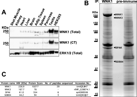
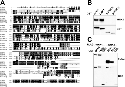
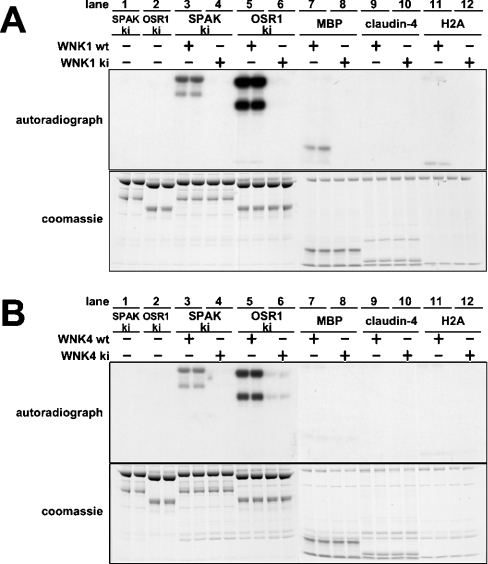
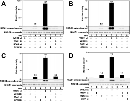
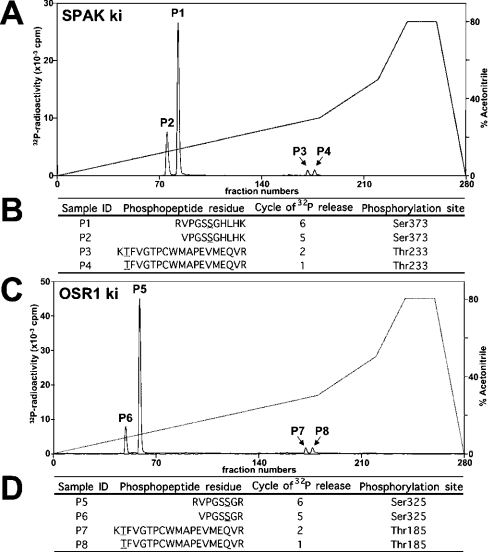
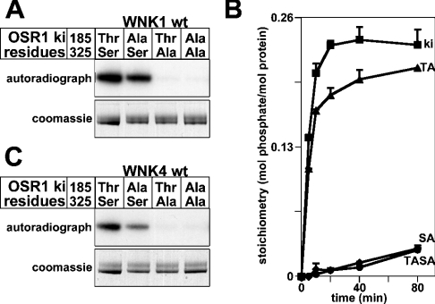
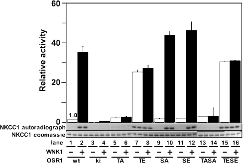
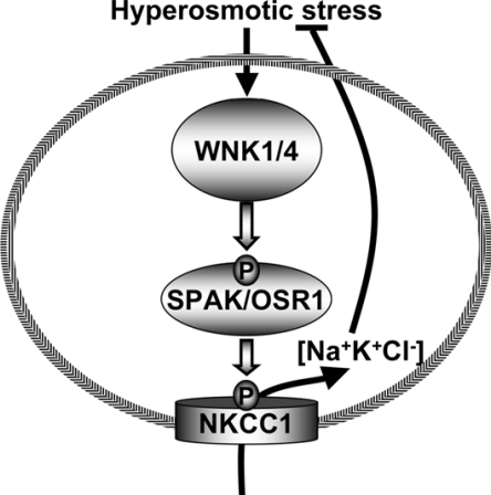
Mutations in the human genes encoding WNK1 [with no K (lysine) protein kinase-1] and the related protein kinase WNK4 are the cause of Gordon's hypertension syndrome. Little is known about the molecular mechanism by which WNK isoforms regulate cellular processes. We immunoprecipitated WNK1 from extracts of rat testis and found that it was specifically associated with a protein kinase of the STE20 family termed 'STE20/SPS1-related proline/alanine-rich kinase' (SPAK). We demonstrated that WNK1 and WNK4 both interacted with SPAK as well as a closely related kinase, termed 'oxidative stress response kinase-1' (OSR1). Wildtype (wt) but not catalytically inactive WNK1 and WNK4 phosphorylated SPAK and OSR1 to a much greater extent than with other substrates utilized previously, such as myelin basic protein and claudin-4. Phosphorylation by WNK1 or WNK4 markedly increased SPAK and OSR1 activity. Phosphopeptide mapping studies demonstrated that WNK1 phosphorylated kinase-inactive SPAK and OSR1 at an equivalent residue located within the T-loop of the catalytic domain (Thr233 in SPAK, Thr185 in OSR1) and a serine residue located within a C-terminal non-catalytic region (Ser373 in SPAK, Ser325 in OSR1). Mutation of Thr185 to alanine prevented the activation of OSR1 by WNK1, whereas mutation of Thr185 to glutamic acid (to mimic phosphorylation) increased the basal activity of OSR1 over 20-fold and prevented further activation by WNK1. Mutation of Ser325 in OSR1 to alanine or glutamic acid did not affect the basal activity of OSR1 or its ability to be activated by WNK1. These findings suggest that WNK isoforms operate as protein kinases that activate SPAK and OSR1 by phosphorylating the T-loops of these enzymes, resulting in their activation. Our analysis also describes the first facile assay that can be employed to quantitatively assess WNK1 and WNK4 activity.


Figures

Figure 1
Figure 1. Analysis of WNK1-binding proteins
(A) Rat tissue extracts (40 μg of protein) were immunoblotted with the indicated antibodies. Similar findings were obtained in at least two separate experiments. (B) Rat testis extracts were subjected to immunoprecipitation with WNK1 or pre-immune antibody. The immunoprecipitates were subjected to PAGE and the protein bands visualized following colloidal Coomassie Blue staining. The major bands observed in the WNK1, but not the pre-immune, purification are indicated. (C) These bands, as well as the corresponding region in the preimmune sample, were excised from the gel, digested in-gel with trypsin, and the identities of the proteins were determined by tryptic-peptide mass-spectral fingerprinting as described in the Materials and methods section. Mascot protein score where a value of >63 is considered significant (P<0.05), number (No.) of peptides sequenced by MALDI-TOF/TOF-MS/MS (SPAK and SDB84) and liquid chromatography–tandem MS (WNK1) and the accession numbers for each protein identified are indicated. Tryptic peptides derived from WNK1, WNK3, SPAK and SDB84 were present in the WNK1, but not in the preimmune, immunoprecipitate.
Figure 2
Figure 2. Interaction of WNK1/WNK4 with SPAK and OSR1
(A) Multiple sequence alignment of the indicated STE20 human kinases. Identical residues are highlighted in black and similar residues are highlighted in grey. The kinase domain is marked with a continuous line, regions of non-similarity between SPAK and OSR1 are marked with a dotted line, the Mg2+-binding aspartic acid residue mutated to inactivate SPAK and OSR1 is marked with a triangle, the T-loop threonine residue phosphorylated by WNK1/WNK4 is marked with an asterisk, and the non-catalytic serine residue phosphorylated by WNK1/WNK4 with a square. (B) HEK-293 cells were transfected with constructs encoding the indicated GST-fusion proteins. At 36 h post-transfection, the GST-fusion proteins were affinity-purified and immunoblotted with GST antibodies or WNK1 antibody to detect endogenously associated WNK1. (C) HEK-293 cells were co-transfected with the indicated combinations of FLAG-tagged WNK1/WNK4, GST–SPAK, GST–OSR1 or empty pEBG2T vector. GST-fusion proteins were affinity-purified and immunoblotted with FLAG antibody to detect WNK1 or WNK4 expression or GST antibody to detect SPAK, OSR1 or GST expression.
Figure 3
Figure 3. Phosphorylation of SPAK/OSR1 by WNK1 and WNK4
(A) Kinase-active wt WNK1-(1–661) or kinase-inactive (ki) [D368A]WNK1-(1–661) was incubated with the indicated proteins (ki-[D212A]SPAK, ki [D164A]OSR1, MBP, claudin-4 and H2A) in the presence of Mg2+ and [γ-32P]ATP. Phosphorylation of protein substrates was determined after PAGE and autoradiography (upper panel) of the Coomassie Blue-stained bands (lower panel) corresponding to each substrate. It should be noted that recombinant SPAK and OSR1 migrate as a doublet band with the upper band migrating with the expected molecular mass for the full-length protein. We presume that the lower band is a proteolytic fragment lacking the C-terminal region, as these enzymes were expressed as N-terminal GST-fusion proteins. (B) As above, expect that kinase-active wt WNK4-(1–593) or ki [K186A/D321A]WNK4-(1–593) were employed. A double mutant of WNK4 was generated in order to ensure complete catalytic inactivation of the kinase. For both (A) and (B) each experimental condition was assayed in duplicate and similar results obtained in at least two different experiments.
Figure 4
Figure 4. Activation of SPAK/OSR1 by WNK1 and WNK4
(A) The indicated combinations of wt or ki WNK1-(1–661) were tested for their ability to activate wt SPAK or ki [D212A]SPAK. Activity of SPAK was assayed employing [1-260]NKCC1 as a substrate. Phosphorylation of NKCC1 was quantified after PAGE and autoradiography (middle panel) of the Coomassie Blue-stained band of NKCC1-(1–260) (lower panel). The phosphorylation of NKCC1 was also measured as 32P radioactivity by Cerenkov counting (upper panel). The results are plotted as means±S.D. for a duplicate experiment relative to the phosphorylation obtained with wt SPAK alone. (B) As above, except that wt OSR1 or ki [D164A]OSR1 were employed. (C and D) As above, except that wt WNK4-(1–593) or ki [K186A/D321A]WNK4-(1–593) were employed.
Figure 5
Figure 5. Analysis of phosphorylation of SPAK and OSR1
(A) ki [D212A]SPAK was phosphorylated by wt WNK1-(1–661) for 40 min under conditions in which phosphorylation was maximal (results not shown). The 32P-labelled SPAK was isolated by PAGE, digested with trypsin and the resulting peptides were chromatographed on a C18 column. Fractions containing the major 32P-labelled peptides are marked. (B) The indicated peptides were analysed by MALDI-TOF/TOF-MS as described in the Materials and methods section. The site of phosphorylation within each peptide was determined by solid-phase Edman sequencing in which 32P radioactivity was measured after each cycle of Edman degradation. The cycle number in which 32P radioactivity was released is indicated. The deduced amino acid sequences of peptides P1, P2, P3, and P4 are indicated; the phosphorylated residues are underlined. (C and D) As above, except that ki [D164A]OSR1 was employed.
Figure 6
Figure 6. Analysis of phosphorylation of OSR1 by WNK1 and WNK4
(A) The indicated mutants of the ki [D164A]OSR1 were phosphorylated by wt WNK1-(1–661) in the presence of Mg2+ and [γ-32P]ATP. Phosphorylation of OSR1 was determined after PAGE and autoradiography (upper panel) of the Coomassie Blue-stained band (lower panel) corresponding to OSR1. (B) As above, except that phosphorylation reactions were terminated at the indicated time points and the stoichiometry of 32P phosphorylation of the indicated mutants of ki OSR1 was determined by Cerenkov counting. The results are plotted as means±S.D. for an experiment performed in duplicate. Abbreviations: ki, kinase-inactive [D164A]OSR1; TA, [D164A/T185A]OSR1; SA, [D164A/S325A]OSR1; TASA, [D164A/T185A/S325A]OSR1. (C) As in (A), except that WNK4-(1–593) was employed.
Figure 7
Figure 7. Analysis of activation of OSR1 by WNK1
The indicated mutants of OSR1 were incubated in the absence (−) or presence (+) of WNK1-(1–661) in the presence of Mg2+ and [γ-32P]ATP. After this incubation, the activity of OSR1 was assayed employing NKCC1-(1–260) as a substrate. Phosphorylation of NKCC1 was quantified after PAGE and autoradiography (middle panel) of the Coomassie Blue-stained band of NKCC1-(1–260) (lower panel). The phosphorylation of NKCC1 was also measured as 32P radioactivity by Cerenkov counting (upper panel). The results are plotted as means±S.D. for a duplicate experiment relative to the phosphorylation obtained with wt OSR1 alone. Abbreviations: ki, kinase-inactive [D164A]OSR1; TA, [T185A]OSR1; TE, [T185E]OSR1; SA, [S325A]OSR1; SE, [S325E]OSR1; TASA, [T185A/S325A]OSR1; TESE, [T185E/S325E]OSR1.
Figure 8
Figure 8. Summary of the mechanism by which hyperosmotic stresses may stimulate NKCC1 co-transporter activity

Comment in

References

    1. Xu B., English J. M., Wilsbacher J. L., Stippec S., Goldsmith E. J., Cobb M. H. WNK1, a novel mammalian serine/threonine protein kinase lacking the catalytic lysine in subdomain II. J. Biol. Chem. 2000;275:16795–16801. - PubMed
    1. Verissimo F., Jordan P. WNK kinases, a novel protein kinase subfamily in multi-cellular organisms. Oncogene. 2001;20:5562–5569. - PubMed
    1. Wilson F. H., Disse-Nicodeme S., Choate K. A., Ishikawa K., Nelson-Williams C., Desitter I., Gunel M., Milford D. V., Lipkin G. W., Achard J. M., et al. Human hypertension caused by mutations in WNK kinases. Science. 2001;293:1107–1112. - PubMed
    1. Zambrowicz B. P., Abuin A., Ramirez-Solis R., Richter L. J., Piggott J., BeltrandelRio H., Buxton E. C., Edwards J., Finch R. A., Friddle C. J., et al. Wnk1 kinase deficiency lowers blood pressure in mice: a gene-trap screen to identify potential targets for therapeutic intervention. Proc. Natl. Acad. Sci. U.S.A. 2003;100:14109–14114. - PMC - PubMed
    1. Kamide K., Takiuchi S., Tanaka C., Miwa Y., Yoshii M., Horio T., Mannami T., Kokubo Y., Tomoike H., Kawano Y., Miyata T. Three novel missense mutations of WNK4, a kinase mutated in inherited hypertension, in Japanese hypertensives: implication of clinical phenotypes. Am. J. Hypertens. 2004;17:446–449. - PubMed

Publication types

MeSH terms