. 2005 Nov 15;392(Pt 1):83-92.
doi: 10.1042/BJ20051102.

Characterization of CA XV, a new GPI-anchored form of carbonic anhydrase

Affiliations

Characterization of CA XV, a new GPI-anchored form of carbonic anhydrase

Mika Hilvo et al. Biochem J. .

Abstract

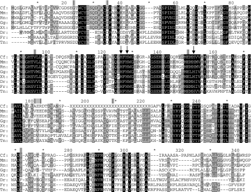
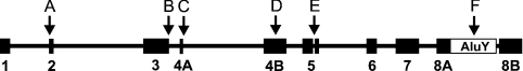
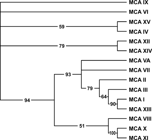
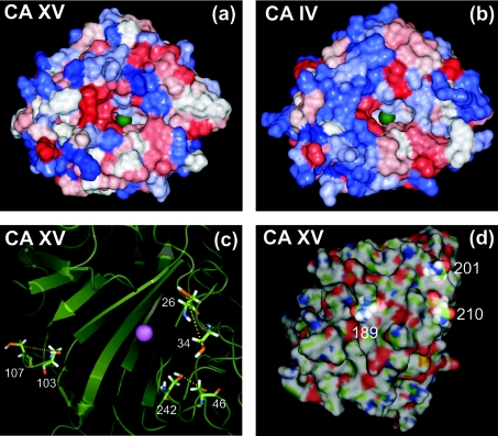
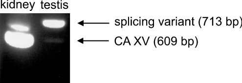
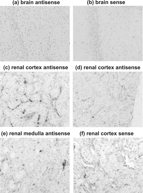
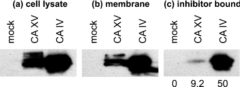
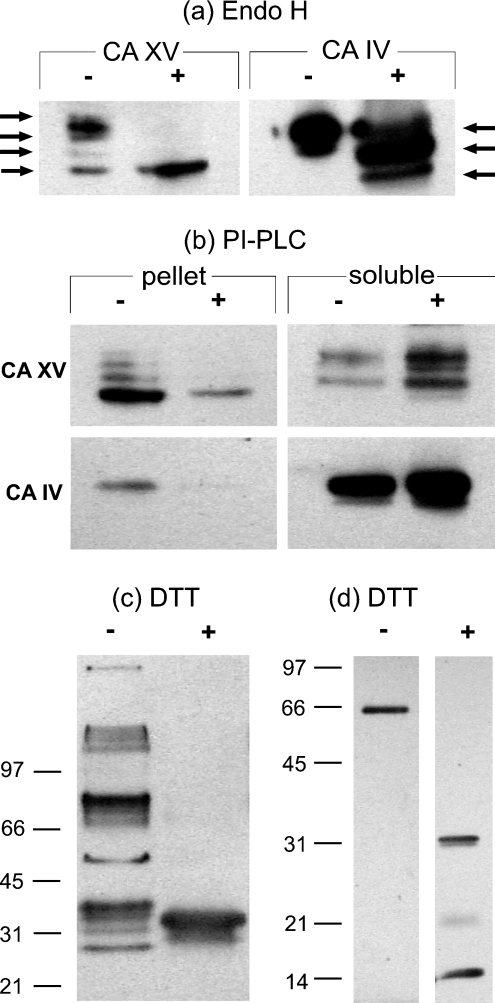
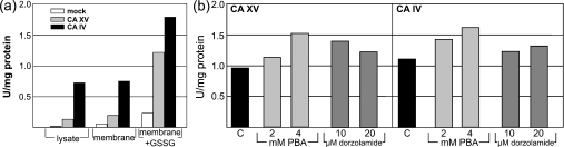
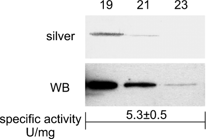
The main function of CAs (carbonic anhydrases) is to participate in the regulation of acid-base balance. Although 12 active isoenzymes of this family had already been described, analyses of genomic databases suggested that there still exists another isoenzyme, CA XV. Sequence analyses were performed to identify those species that are likely to have an active form of this enzyme. Eight species had genomic sequences encoding CA XV, in which all the amino acid residues critical for CA activity are present. However, based on the sequence data, it was apparent that CA XV has become a non-processed pseudogene in humans and chimpanzees. RT-PCR (reverse transcriptase PCR) confirmed that humans do not express CA XV. In contrast, RT-PCR and in situ hybridization performed in mice showed positive expression in the kidney, brain and testis. A prediction of the mouse CA XV structure was performed. Phylogenetic analysis showed that mouse CA XV is related to CA IV. Therefore both of these enzymes were expressed in COS-7 cells and studied in parallel experiments. The results showed that CA XV shares several properties with CA IV, i.e. it is a glycosylated glycosylphosphatidylinositol-anchored membrane protein, and it binds CA inhibitor. The catalytic activity of CA XV is low, and the correct formation of disulphide bridges is important for the activity. Both specific and non-specific chaperones increase the production of active enzyme. The results suggest that CA XV is the first member of the alpha-CA gene family that is expressed in several species, but not in humans and chimpanzees.


Figures

Figure 1
Figure 1. The results of sequence analyses
The alignment of CA XV in eight species, showing its conservation throughout evolution. The three histidine residues co-ordinating the zinc atom are pointed out by arrows. Exon boundaries are indicated by vertical lines above the alignment; asterisks denote every tenth residue not labelled in the Figure. The only exon boundary having interspecies differences is located at amino acid residues 181–183. Abbreviations: Cf, Canis familiaris; Mm, Mus musculus; Rn, Rattus norvegicus; Gg, Gallus gallus; Xt, Xenopus tropicalis; Dr, Danio rerio; Fr, Fugu rubripes; Tn, Tetraodon nigroviridis. X, sequencing gap in the genome.
Figure 2
Figure 2. Organization of human and chimpanzee CA XV pseudogenes
Numbered black boxes show reconstructed exons. Exon and intron lengths are presented in the correct scale. Arrows highlight those defects that are common to all five copies. Arrows A and B: frame-shifts. Arrow C, the beginning of the intron after exon 4A has GA instead of the conserved GT dinucleotide. Arrow D, a 9 bp insertion in exon 4B, disrupting the active centre. Arrow E, a 4-bp insertion in exon 5, leading to a frame-shifted sequence in a region which is highly conserved in all CAs. Arrow F, insertion of an AluY-repeat sequence which splits exon 8, duplicating 17 bp of the exon sequence (duplicated part seen as a gap after AluY).
Figure 3
Figure 3. The phylogenetic tree of 15 mouse CAs and CA-RPs
The numbers at the branches show confidence levels in the bootstrap analysis. The tree implies that mouse CA XV is most closely related to CA IV.
Figure 4
Figure 4. The prediction of the structure for CA XV
Panels (a) and (b): comparison of the surfaces of CA XV and CA IV, coloured according to hydropathy (blue represents hydrophilic, and red hydrophobic). The green sphere represents the zinc atom crucial for CA activity. Panel (c): secondary structures and cysteine pairs. The zinc atom is shown by a purple sphere. Panel (d): highlighted asparagines predicted to represent glycosylation sites; residue numbers are the same as shown as in Figure 1.
Figure 5
Figure 5. Results of RT-PCR
The results of the kidney and testis RT-PCR are shown in the Figure. The kidney has a strong band for CA XV (609 bp), whereas in the testis the band is much weaker. In testis, however, there is a stronger band for a splicing variant (713 bp). The rest of the results are summarized in Table 1.
Figure 6
Figure 6. In situ hybridization revealing CA15 mRNA in mouse tissues
The signal was present in brain (a), whereas the negative control had a much lower signal density (b). High expression was observed in the renal cortex (c, d) and lower expression in the medulla (e). The sense control shows the background level of the signal (f). Original magnifications of the panels: a, b, c, e and f, ×400; d, ×200.
Figure 7
Figure 7. Expression of functional mouse CA XV in the membrane of transfected COS-7 cells
(a) Total cell lysates of non-transfected COS-7 cell (mock) and transfected COS-7 cells (with CA15 or CA4 cDNAs) were analysed by Western blotting using anti-mouse CA IV antibodies. (b) The cell lysates were centrifuged at 100000 g for 30 min. The membranes were analysed by Western blotting. (c) The membrane extracts in 10 mM Hepes/NaOH, pH 7.5, containing 1% NP40 and protease inhibitors were incubated with CA inhibitor-affinity resin, and bound enzyme was analysed by Western blotting. The numbers at the bottom of the gel show percentages of inhibitor-bound CA XV or CA IV.
Figure 8
Figure 8. Biochemical properties of mouse CA XV
(a) EndoH treatment resulted in shifts of high-molecular-mass polypeptide of CA XV and CA IV. The CA XV showed two intermediate polypeptides, whereas CA IV showed only one intermediate polypeptide, as indicated by arrows. (b) COS-7 cell membranes were treated without (−) and with (+) PI-PLC enzyme. The enzymes remaining in the membrane pellet and soluble fraction after PI-PLC treatment were analysed by Western blotting. The decrease in the membrane-associated CA XV was compensated by an increase in the soluble fraction after PI-PLC treatment, suggesting that CA XV is a GPI-anchored protein. (c) COS-7 cell lysates expressing mouse CA XV were analysed by Western blotting under non-reducing (−) and reducing (+) conditions. Under non-reducing conditions, the mouse CA XV formed high-molecular-mass aggregates, which upon reducing with DTT resulted in a single immunoreactive polypeptide. (d) Affinity pure recombinant mouse CA XV from E. coli was treated with non-reducing (−) and reducing (+) sample buffer and analysed by SDS/PAGE, followed by Western blotting.
Figure 9
Figure 9. Refolding of CA XV by oxidized glutathione and chemical chaperones
(a) The cell lysates of COS-7 cells transfected with CA15 or CA4 cDNAs and untransfected cells (mock) were analysed for CA activity. The membrane suspensions from each cell line were also used for CA activity measurement. The results suggested that the cell membranes contained detectable CA XV and IV activities. Upon treatment with 10 mM GSSG, both CA XV and CA IV were refolded further into more active enzymes. (b) The COS-7 cells, after transfection with CA15 or CA4 cDNAs, were treated with 2 and 4 mM PBA or 10 and 20 μM dorzolamide for 72 h. After removing the PBA or dorzolamide, the cell membranes were used for CA activity measurement. Both non-specific (PBA) and specific (dorzolamide) chemical chaperones were able to refold the mouse CA XV and IV into more active enzymes. Abbreviation: U, units.
Figure 10
Figure 10. Recombinant affinity-purified mouse CA XV from E. coli
The fractions from the Sephacryl S-300 sizing column were analysed by SDS/PAGE. The polypeptides were visualized by silver staining (silver) or Western blot analysis (WB). The numbers show the fractions used for SDS/PAGE. The homogeneous pooled enzyme showed a specific activity of 5.3±0.5 units/mg.

References

    1. Lindskog S., Silverman D. N. The catalytic mechanism of mammalian carbonic anhydrases. In: Chegwidden W. R., Carter N. D., Edwards Y. H., editors. The Carbonic Anhydrases: New Horizons. Basel: Birkhäuser Verlag; 2000. pp. 175–195. - PubMed
    1. Sly W. S., Hu P. Y. Human carbonic anhydrases and carbonic anhydrase deficiencies. Annu. Rev. Biochem. 1995;64:375–401. - PubMed
    1. Hewett-Emmett D. Evolution and distribution of the carbonic anhydrase gene families. In: Chegwidden W. R., Carter N. D., Edwards Y. H., editors. The Carbonic Anhydrases: New Horizons. Basel: Birhkhäuser Verlag; 2000. pp. 29–76. - PubMed
    1. Lehtonen J., Shen B., Vihinen M., Casini A., Scozzafava A., Supuran C. T., Parkkila A. K., Saarnio J., Kivela A. J., Waheed A., et al. Characterization of CA XIII, a novel member of the carbonic anhydrase isoenzyme family. J. Biol. Chem. 2004;279:2719–2727. - PubMed
    1. Fleming R. E., Parkkila S., Parkkila A. K., Rajaniemi H., Waheed A., Sly W. S. Carbonic anhydrase IV expression in rat and human gastrointestinal tract regional, cellular, and subcellular localization. J. Clin. Invest. 1995;96:2907–2913. - PMC - PubMed

Publication types

MeSH terms