

Integration of engrafted Schwann cells into injured peripheral nerve: axonal association and nodal formation on regenerated axons
- PMID: 16084645
- PMCID: PMC2605373
- DOI: 10.1016/j.neulet.2005.06.073
Integration of engrafted Schwann cells into injured peripheral nerve: axonal association and nodal formation on regenerated axons
Abstract
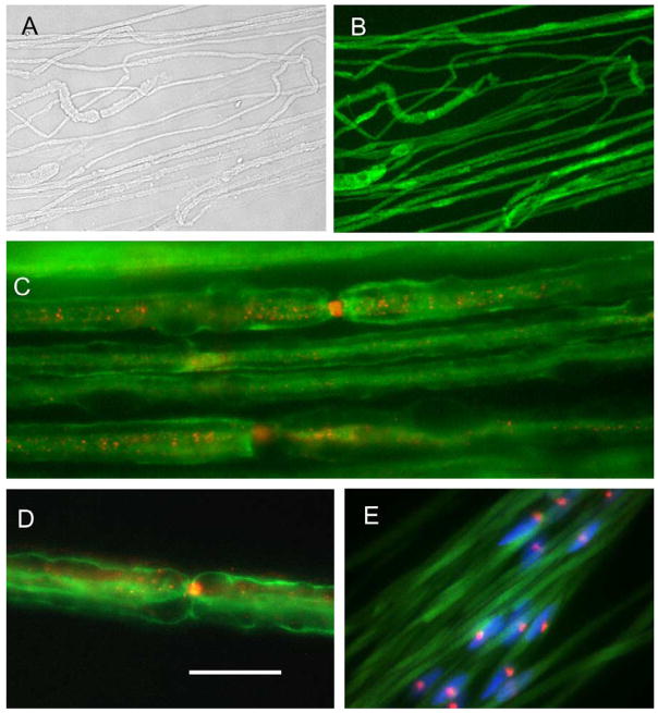
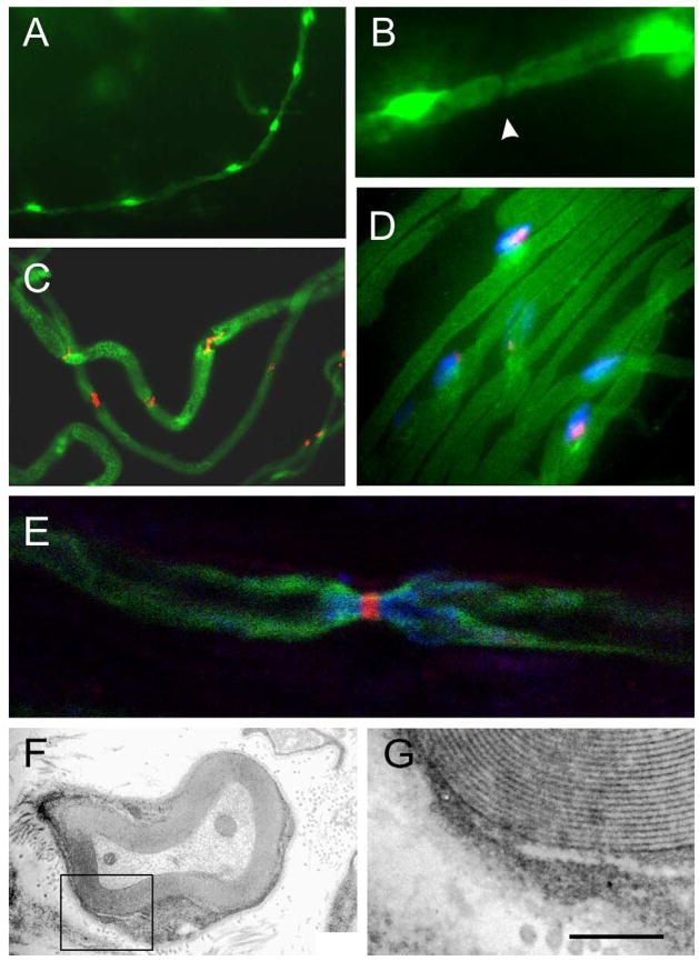
Transplantation of myelin-forming cells can remyelinate axons, but little is known of the sodium channel organization of axons myelinated by donor cells. Sciatic nerve axons of female wild type mice were transected by a crush injury and Schwann cells (SCs) from green fluorescence protein (GFP)-expressing male mice were transplanted adjacent to the crush site. The male donor cells were identified by GFP fluorescence and fluorescence in situ hybridization (FISH) for Y chromosome. In nerves of GFP-expressing mice, GFP was observed in the axoplasm and in the cytoplasmic compartments of the Schwann cells, but not in the myelin. Following transplantation of GFP-SCs into crushed nerve of wild type mice, immuno-electron microscopic analysis indicated that GFP was observed in the cytoplasmic compartments of engrafted Schwann cells which formed myelin. Nodal and paranodal regions of the axons myelinated by the GFP-SCs were identified by Na(v)1.6 sodium channel and Caspr immunostaining, respectively. Nuclear identification of the Y chromosome by FISH confirmed the donor origin of the myelin-forming cells. These results indicate that engrafted GFP-SCs participate in myelination of regenerated peripheral nerve fibers and that Na(v)1.6 sodium channel, which is the dominant sodium channel at normal nodes, is reconstituted on the regenerated axons.
Figures
References
-
- Baron-Van Evercooren A, Gansmuller A, Duhamel E, Pascal F, Gumpel M. Repair of a myelin lesion by Schwann cells transplanted in the adult mouse spinal cord. Neuroimmunology. 1992;40:235–242. - PubMed
-
- Berthold C-H. Morphology of normal peripheral axons. In: Waxman SG, editor. Physiology and Pathobiology of Axons. Raven Press; New York: 1978. pp. 3–64.
-
- Blakemore WF. Remyelination of CNS axons by Schwann cells transplanted from the sciatic nerve. Nature. 1977;266:68–69. - PubMed
-
- Duncan ID, Hammang JP, Jackson KF, Wood PM, Bunge RP, Langford L. Transplantation of oligodendrocytes and Schwann cells into the spinal cord of the myelin-deficient rat. J Neurocytol. 1988;17:351–360. - PubMed
Publication types
MeSH terms
Substances
Grants and funding
LinkOut - more resources
Full Text Sources
Medical
