. 2005 Aug 5;33(14):4412-24.
doi: 10.1093/nar/gki747. Print 2005.

Initiation of DNA replication at the human beta-globin 3' enhancer

Affiliations

Initiation of DNA replication at the human beta-globin 3' enhancer

Alla Buzina et al. Nucleic Acids Res. .

Abstract

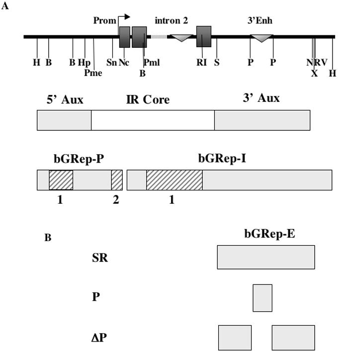
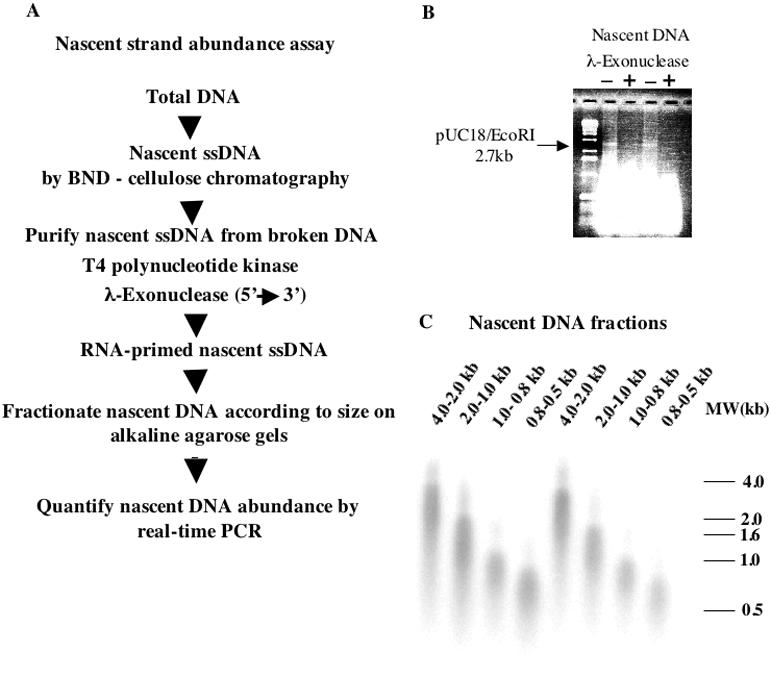
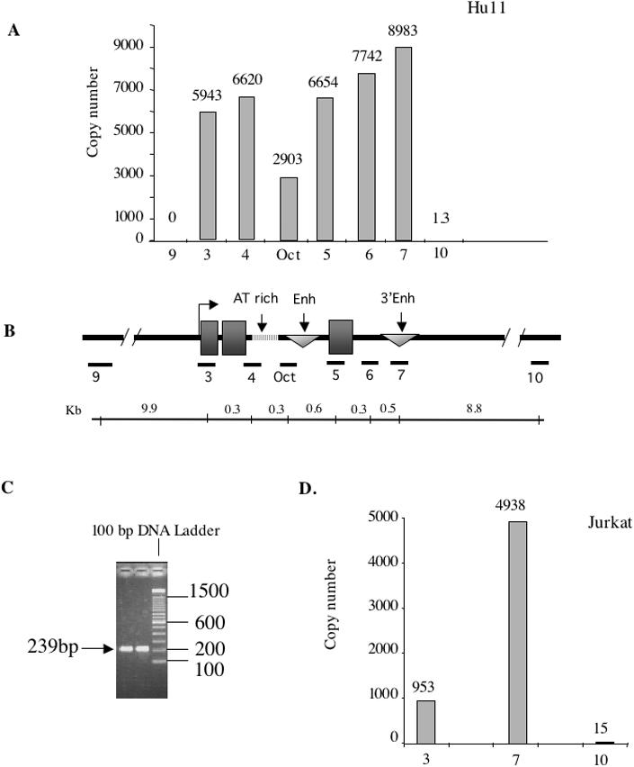
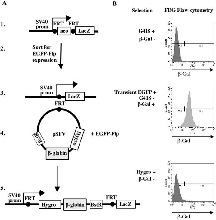
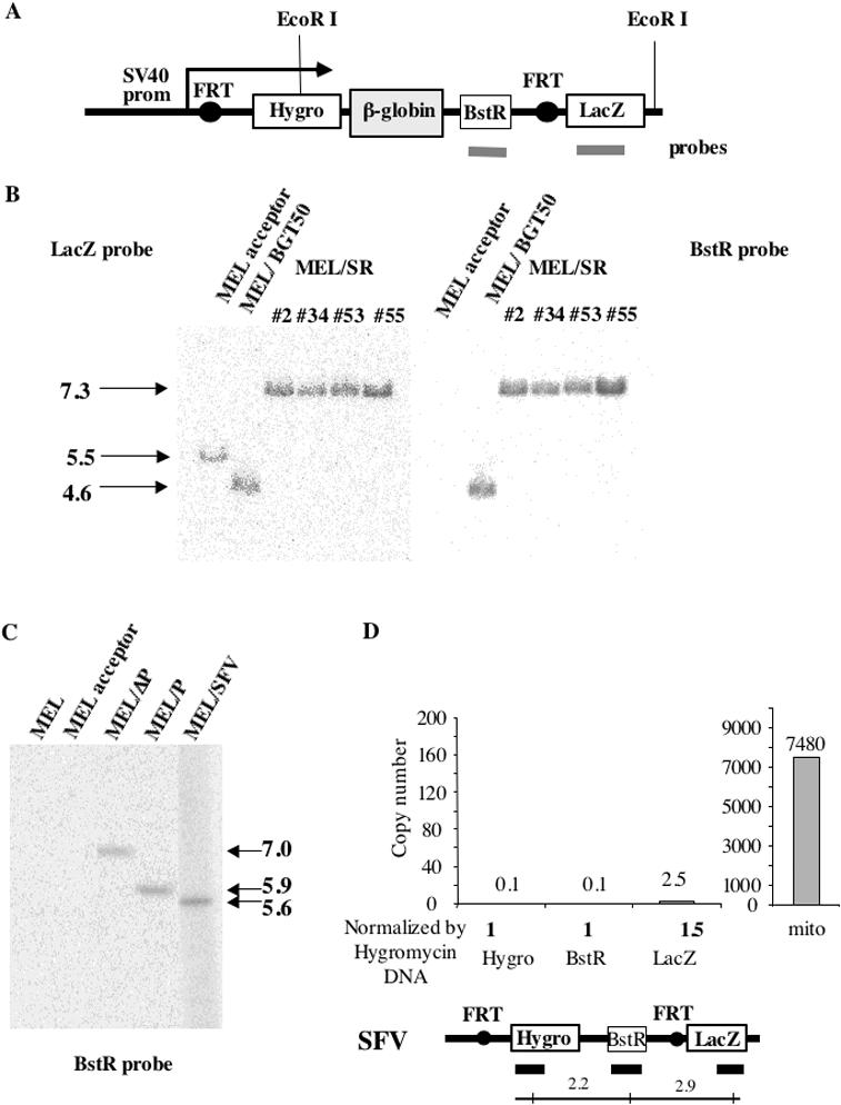
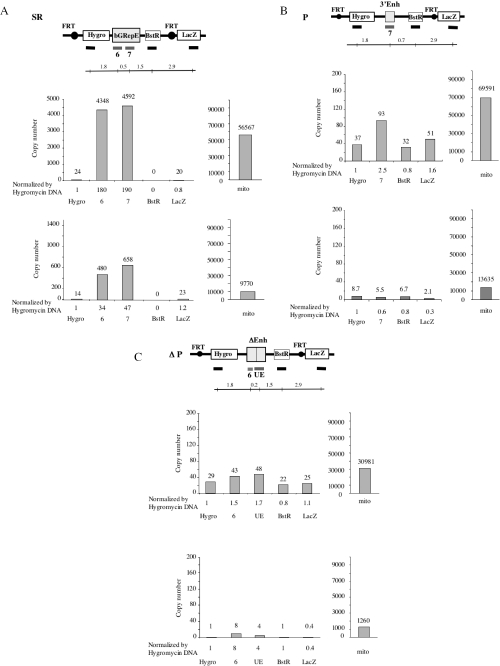
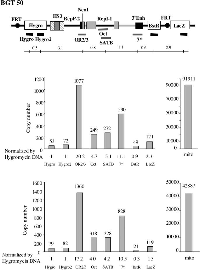
The origin of DNA replication in the human beta-globin gene contains an initiation region (IR) and two flanking auxiliary elements. Two replicator modules are located within the upstream auxiliary sequence and the IR core, but the functional sequences in the downstream auxiliary element are unknown. Here, we use a combination of benzoylated-naphthoylated DEAE (BND) cellulose purification and nascent strand abundance assays to show that replication initiation occurs at the beta-globin 3' enhancer on human chromosome 11 in the Hu11 hybrid murine erythroleukemia (MEL) cell line. To examine replicator function, 3' enhancer fragments were inserted into an ectopic site in MEL cells via an optimized FRT/EGFP-FLP integration system. These experiments demonstrate that the 1.6 kb downstream auxiliary element is a third replicator module called bGRep-E in erythroid cells. The minimal 260 bp 3' enhancer is required but not sufficient to initiate efficient replication, suggesting cooperation with adjacent sequences. The minimal 3' enhancer also cooperates with elements in an expressing HS3beta/gamma-globin construct to initiate replication. These data indicate that the beta-globin replicator has multiple initiation sites in three closely spaced replicator modules. We conclude that a mammalian enhancer can cooperate with adjacent sequences to create an efficient replicator module.


Figures

Figure 1
Figure 1
Map of the human β-globin gene and replication origin. (A) Solid boxes represent β-globin exons, triangles are β-globin enhancers, the banded line is an AT-rich element. Restriction sites: B-BamHI, H-HindIII, Hp-HpaI, N-NdeI, Nc-NcoI, P-PstI, RI-EcoRI, RV-EcoRV, S-SwaI, Sn-SnaBI, X-XbaI. IR core – initiation region core; 5′ and 3′ Aux – upstream and downstream auxiliary sequences; bGRep-P and bGRep-I known replicator modules with essential subregions shown as hatched boxes. (B) β-globin replicator fragments used in this study for fine mapping replication initiation in the downstream auxiliary sequence.
Figure 2
Figure 2
Replication initiation assay. (A) Schematic representation of the methodology used for the nascent strand abundance assay. (B) Quality assessment of enzymatic reactions by adding linearized plasmid DNA to the nascent DNA samples. Plasmid DNA has been completely digested after phosphorylation and λ-exonuclease treatment, which indicates that broken DNA has been removed from the nascent DNA samples. (C) Southern blot analysis of the nascent DNA fractions probed with labeled genomic DNA from Hu11 cells to verify DNA fractionation. The 0.8–1 kb nascent DNA fraction was subjected to abundance analysis by real-time PCR.
Figure 3
Figure 3
Replication initiation profile of the endogenous β-globin gene. (A) Real-time PCR analysis of nascent DNA from Hu11 cells. Primer pairs cover the entire β-globin gene including the promoter and 3′ enhancer. Control primer pairs localized 10 kb upstream and 9 kb downstream of the β-globin gene did not detect any replication initiation activity (primer pairs 9 and 10). Primer pair 3 revealed nascent DNA abundance near the β-globin core promoter as expected (3,4). Nascent strand abundance peaks at primer pair 7 in the 3′ enhancer suggesting the presence of another replicator. Copy number is the absolute value obtained from a sample dilution that produces linear amplification. (B) Structure of the β-globin gene with the distance between chosen primer pairs shown under the map. (C) Electrophoresis of final PCR products amplified with primer pair 7. (D) Real-time PCR data of replication initiation activity at the β-globin gene in nascent DNA obtained by the sucrose gradient method from the Jurkat human T-cell leukemia cell line. Primer pair 7 revealed abundant amplification at the 3′ enhancer, indicating initiation of replication.
Figure 4
Figure 4
Site-specific integration of globin sequences into acceptor MEL cells. (A) Schematic representation of the steps involved in obtaining the MEL acceptor cell line with the single FRT site and integration of the globin sequences at this site. (1) Stable transfection of vector DNA containing two FRT sites. (2) Transient expression of EGFP-FLP to excise the Neo gene by FLP-mediated recombination at FRT sites. (3) Structure of the MEL acceptor cell line bearing a single FRT site. (4) Transient co-transfection of SFV/globin cassette and EGFP-FLP vector to integrate globin sequence at the FRT site. (5) Structure of the integrated globin cassette at the FRT site in MEL cells. (B) Selection strategy. (1) Stable transfection of MEL cells resulted in G418 resistant clones. Due to interruption of the SV40 promoter and LacZ sequence by the Neo gene these clones were β-gal negative as detected by FDG Flow cytometry. (2) Transient expression of EGFP-FLP protein allowed enrichment for cells expressing FLP recombinase by Flow sorting of EGFP-positive cells. (3) FLP-mediated recombination at FRT sites excised the Neo sequence and brought LacZ gene under the control of the SV40 promoter. β-gal positive clones were selected by FDG staining and Flow cytometry. (4) Hygromycin resistant clones, in which integration of SFV/globin cassettes occurred, were selected, expanded and stained with FDG to select for clones with FLP-mediated integration at the FRT site.
Figure 5
Figure 5
Single copy intact integration of globin sequences at the fixed FRT site. (A) Structure of targeted MEL cells. Genomic DNA was digested with EcoRI. Southern Blots were hybridized with labeled LacZ or BstR DNA probes (grey lines). (B) Digestion of genomic DNA with EcoRI (which cuts the integrated vector DNA once and cuts MEL acceptor DNA 3′ of LacZ) hybridized with LacZ probe (left) confirming intactness of the integrated constructs. Hybridization with BstR probe detected no band in the MEL acceptor cell line as expected and a single band of the expected size indicating single site integration of the β-globin sequences at the FRT site and the absence of randomly integrated vector DNA. (C) Southern analysis for single site integration in additional constructs produces the correct size bands after hybridization with the BstR probe. (D) Real-time PCR analysis of nascent DNA from MEL/SFV cells indicating that there is no background replication initiation activity at LacZ, BstR and Hygro marker gene sequences. Abundance of amplification with primers specific to mitochondrial DNA indicates the quality of the nascent DNA preparations.
Figure 6
Figure 6
The downstream auxiliary sequence initiates replication at the ectopic site. (A) Genomic structure of MEL/SR cells and real-time PCR quantification data of two independent MEL/SR nascent DNA preparations. A peak of initiation at the 3′ enhancer (primer pair 7) suggests the existence of a third replicator module called bGRep-E. Abundance is presented as mean number of molecules (copy number) in duplicate samples for each nascent preparation obtained using a dilution of the sample that produces linear amplification. The distance between chosen primer pairs is shown under the map. (B) Genomic structure of MEL/P cells and real-time PCR quantification data of two independent MEL/P nascent DNA preparations. Absence of specific amplification with primer pair 7 despite the high abundance of the mitochondrial DNA in the two nascent DNA preparations, indicates that the minimal 260 bp 3′ enhancer sequence is not sufficient to initiate efficient replication. (C) Genomic structure of MEL/ΔP cells and real-time PCR analysis on duplicate nascent DNA preparations using primers 5′ of the PstI deletion that detected only background amplification. These data suggest that deletion of the minimal 3′enhancer abolishes initiation of replication on surrounding sequences.
Figure 7
Figure 7
Initiation at the minimal 3′ enhancer in an HS3 β/γ globin cassette. Genomic structure of MEL/BGT50 cells and real-time PCR quantification data of two independent MEL/BGT50 nascent DNA preparations showing two peaks of DNA replication initiation at the 3′ enhancer (primer pair 7*) and near the globin proximal promoter (primer pair OR2/3). The distance between chosen primer pairs is shown under the map. These data suggest that downstream auxiliary sequences in the bGRep-E can be compensated for by HS3 or the proximal promoter (bGRep-P-2) or intron 2 (bGRep-I-1).

Similar articles

Cited by

References

    1. Kitsberg D., Selig S., Keshet I., Cedar H. Replication structure of the human beta-globin gene domain. Nature. 1993;366:588–590. - PubMed
    1. Aladjem M.I., Groudine M., Brody L.L., Dieken E.S., Fournier R.E., Wahl G.M., Epner E.M. Participation of the human beta-globin locus control region in initiation of DNA replication. Science. 1995;270:815–819. - PubMed
    1. Aladjem M.I., Rodewald L.W., Kolman J.L., Wahl G.M. Genetic dissection of a mammalian replicator in the human beta-globin locus. Science. 1998;281:1005–1009. - PubMed
    1. Cimbora D.M., Schubeler D., Reik A., Hamilton J., Francastel C., Epner E.M., Groudine M. Long-distance control of origin choice and replication timing in the human beta-globin locus are independent of the locus control region. Mol. Cell. Biol. 2000;20:5581–5591. - PMC - PubMed
    1. Pasceri P., Pannell D., Wu X., Ellis J. Full activity from human beta-globin locus control region transgenes requires 5′HS1, distal beta-globin promoter, and 3′ beta-globin sequences. Blood. 1998;92:653–663. - PubMed

Publication types