

1,2-diacyl-phosphatidylcholine flip-flop measured directly by sum-frequency vibrational spectroscopy
- PMID: 16085770
- PMCID: PMC1366751
- DOI: 10.1529/biophysj.105.065672
1,2-diacyl-phosphatidylcholine flip-flop measured directly by sum-frequency vibrational spectroscopy
Abstract
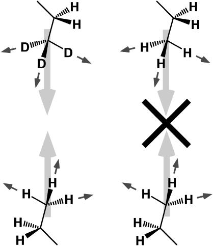
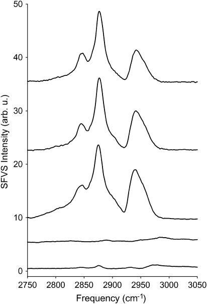
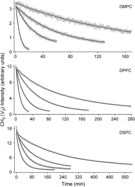
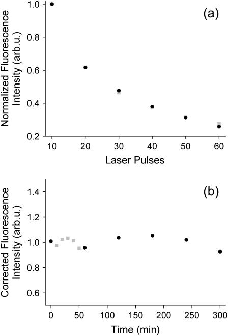
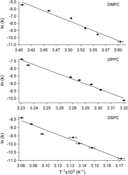
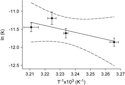
Sum-frequency vibrational spectroscopy (SFVS) is used to measure the intrinsic rate of lipid flip-flop for 1,2-dimyristoyl-sn-glycero-3-phosphocholine (DMPC), 1,2-dipalmitoyl-sn-glycero-3-phosphocholine (DPPC), and 1,2-distearoyl-sn-glycero-3-phosphocholine (DSPC) in planar-supported lipid bilayers (PSs). Asymmetric PSLBs were prepared using the Langmuir-Blodgett/Langmuir-Schaefer method by placing a perdeuterated lipid analog in one leaflet of the PSLB. SFVS was used to directly measure the asymmetric distribution of the native lipid within the membrane by measuring the decay in the CH3 v(s) intensity at 2875 cm(-1) with time and as a function of temperature. An average activation energy of 220 kJ/mol for the translocation of DMPC, DPPC, and DSPC was determined. A decrease in alkyl chain length resulted in a substantial increase in the rate of flip-flop manifested as an increase in the Arrhenius preexponential factor. The effect of lipid labeling was investigated by measuring the exchange of 1,2-dipalmitoyl-sn-glycero-3-phosphoethanolamine-n,n-Dimethyl-n-(2',2',6',6'-tetramethyl-4'-piperidyl) (TEMPO-DPPC). The rate of TEMPO-DPPC flip-flop was an order-of-magnitude slower compared to DPPC. An activation energy of 79 kJ/mol was measured which is comparable to that previously measured by electron spin resonance. The results of this study illustrate how SFVS can be used to directly measure lipid flip-flop without the need for a fluorescent or spin-labeled lipid probe, which can significantly alter the rate of lipid translocation.
Figures
References
-
- Kol, M. A., B. de Kruijff, and A. I. P. M. de Kroon. 2002. Phospholipid flip-flop in biogenic membranes: what is needed to connect opposite sides. Semin. Cell Dev. Biol. 13:163–170. - PubMed
-
- Buton, X., G. Morrot, P. Fellmann, and M. Seigneuret. 1996. Ultra-fast glycerophospholipid selective transbilayer motion mediated by a protein in the endoplasmic reticulum membrane. J. Biol. Chem. 271:6651–6658. - PubMed
-
- Herrmann, A., A. Zachowski, and P. F. Devaux. 1990. Protein mediated phospholipid translocation in the endoplasmic reticulum with a low lipid specificity. Biochemistry. 29:2023–2027. - PubMed
-
- Martin, O. C., and R. E. Pagano. 1987. Transbilayer movement of fluorescent analogs of phosphatidylserine and phosphatidylethanolamine at the plasma membrane of cultured cells. Evidence for a protein-mediated and ATP-dependent process(es). J. Biol. Chem. 262:5890–5898. - PubMed
-
- Eytan, G. D., R. Regev, G. Oren, and Y. G. Assaraf. 1996. The role of passive transbilayer drug movement in multidrug resistance and its modulation. J. Biol. Chem. 271:12897–12902. - PubMed
Publication types
MeSH terms
Substances
Grants and funding
LinkOut - more resources
Full Text Sources
