. 2005 Nov;89(5):3042-8.
doi: 10.1529/biophysj.105.067124. Epub 2005 Aug 5.

Cav1.4 encodes a calcium channel with low open probability and unitary conductance

Affiliations

Cav1.4 encodes a calcium channel with low open probability and unitary conductance

Clinton J Doering et al. Biophys J. 2005 Nov.

Abstract

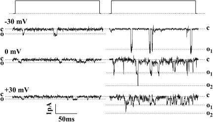
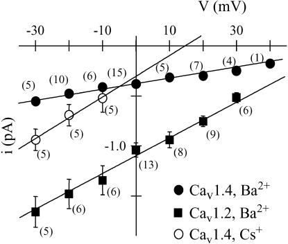
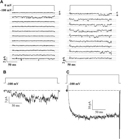
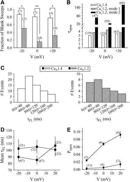
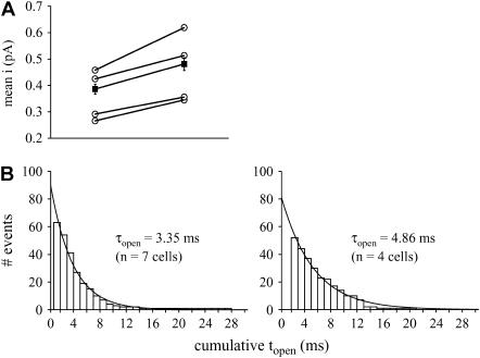
When transiently expressed in tsA-201 cells, Ca(v)1.4 calcium channels support only modest whole-cell currents with unusually slow voltage-dependent inactivation kinetics. To examine the basis for this unique behavior we used cell-attached patch single-channel recordings using 100 mM external barium as the charge carrier to determine the single-channel properties of Ca(v)1.4 and to compare them to those of the Ca(v)1.2. Ca(v)1.4 channel openings occurred infrequently and were of brief duration. Moreover, openings occurred throughout the duration of the test depolarization, indicating that the slow inactivation kinetics observed at the whole-cell level are caused by sustained channel activity. Ca(v)1.4 and Ca(v)1.2 channels displayed similar latencies to first opening. Because of the rare occurrence of events, the probability of opening could not be precisely determined but was estimated to be <0.015 over a voltage range of -20 to +20 mV. The single-channel conductance of Ca(v)1.4 channels was approximately 4 pS compared with approximately 20 pS for Ca(v)1.2 under the same experimental conditions. Additionally, in the absence of divalent cations, Ca(v)1.4 channels pass cesium ions with a single-channel conductance of approximately 21 pS. Although Ca(v)1.2 opening events were best described kinetically with two open time constants, Ca(v)1.4 open times were best described by a single time constant. BayK8644 slightly enhanced the single-channel conductance in addition to increasing the open time constant for Ca(v)1.4 channels by approximately 45% without, however, causing the appearance of an additional slower gating mode. Overall, our data indicate that single Ca(v)1.4 channels support only minute amounts of calcium entry, suggesting that large numbers of these channels are needed to allow for significant whole-cell current activity, and providing a mechanism to reduce noise in the visual system.


Figures

FIGURE 1
FIGURE 1
Single-channel openings observed for Cav1.4 (left) and Cav1.2 channels (right) when cotransfected with ancillary α2-δ1 and β2a subunits. The events were recorded with 100 mM barium as the charge carrier in the absence of agonist BayK8644. The events were observed by depolarizing from a holding potential (physiological) of −100 mV to −30 mV (top traces), 0 mV (middle traces), and +30 mV (bottom traces). Dashed lines denote closed (c) and open (o) levels. Coincident openings are evident for Cav1.2 indicating that at least two channels were in the patch. Note that Cav1.4 channel openings are smaller in amplitude and occur less frequently than those observed with Cav1.2. Both records were filtered at the same frequency; the difference in noise is due to recordings having been performed at different times when the root mean-square noise differed.
FIGURE 2
FIGURE 2
Current-voltage relations for single Cav1.4 (circles) and Cav1.2 (squares) channels, with either barium (solid symbols) or cesium (open symbols) as the charge carrier. Data points are average event amplitudes mean ± SE, and numbers in parentheses indicate number of cells (note that error bars for Cav1.4 data are too small to be visible). Linear regression indicates that the unitary conductance is 3.7 ± 0.3 pS for Cav1.4 and 19.5 ± 0.1 pS for Cav1.2 in barium, and 21 ± 2 pS for Cav1.4 in cesium. The observed values are statistically significantly different (p < 0.05).
FIGURE 3
FIGURE 3
(A) Example of consecutive sweeps for Cav1.4 (left) and Cav1.2 (right). Note that events occur in relatively few traces with Cav1.4 and in most traces with Cav1.2. Traces were filtered at the same frequencies. (B) Ensemble current obtained by pooling data from three cells (600 sweeps, 39 of which showed events). Cells were from the same transfection. (C) Whole-cell current recorded in 100 mM external barium from the same transfection as the data shown in B. The test potential was +40 mV.
FIGURE 4
FIGURE 4
Analysis of single channel kinetics. (A) Fraction of blank sweeps for Cav1.4 and Cav1.2, at three different voltages. For a given cell, the number of sweeps containing no events was divided by the total number of sweeps (in this case, the total number was determined to be the sweep number in which the last event was observed), and this fraction averaged among different cells (indicated in parentheses). (B) Mean open times for Cav1.4 and Cav1.2 at three different test potentials determined from cumulative open time histograms. Cav1.4 events were best described by a single exponential fit, whereas Cav1.2 events required biexponential fits. Numbers in parentheses indicate total numbers of events in the open time histograms from which τopen values were determined (Cav1.4, 15 cells; Cav1.2, 13 cells). (C) Histograms showing latency to first opening (tFL) for Cav1.4 (left) and Cav1.2 (right) channels at −20 mV. Both Cav1.4 and Cav1.2 show similar first latency profiles. The bin width was 40 ms. (D) Mean first latency times for Cav1.4 and Cav1.2 channels. There was no statistical difference between the two channels. (E) Open probability of Cav1.4 channels determined from the ratio of total open time to total time for a given patch. Note that the open probability for Cav1.4 is more than 10-fold lower than for Cav1.2 channels (all values statistically significant, p < 0.001). Number in parentheses denotes number of cells.
FIGURE 5
FIGURE 5
(A) Application of 10-μM BayK8644 increases single-channel amplitude at +20 mV. Hollow symbols indicate average data from individual cells, and the solid symbols indicate mean values with standard errors. Data in the presence and the absence of BayK were obtained from the same cells. BayK significantly increases the mean current from 0.39 ± 0.02 pA (n = 26 events) to 0.48 ± 0.02 pA (n = 52 events) (p < 0.05; paired Student's t-test). (B) BayK increases mean open time, τopen. Cumulative open time histograms are shown in the absence (left) and presence (right) of BayK for events observed at +20 mV. The data are best described by a monoexponential fit (r2 = 0.99 each). Here, the control data include cells that were not subsequently exposed to BayK.
FIGURE 6
FIGURE 6
Alignment of p-loop regions of L-type calcium channels. Note that only one residue differs in each of domains I, III, and IV among the four family members, highlighted in bold. Glutamate residues thought to be important for selectivity are shown highlighted. Given the high sequence homology between Cav1.2 and Cav1.4, and the dramatically different unitary conductances and open probabilities, it is likely that residues outside of the pore region are important determinants in permeation.

References

    1. Schneeweis, D. M., and J. L. Schnapf. 1995. Photovoltage of rods and cones in the macaque retina. Science. 268:1053–1056. - PubMed
    1. Taylor, W. R., and C. Morgans. 1998. Localization and properties of voltage-gated calcium channels in cone photoreceptors of Tupaia belangeri. Vis. Neurosci. 15:541–552. - PubMed
    1. Corey, D. P., J. M. Dubinsky, and E. A. Schwartz. 1984. The calcium current in inner segments of rods from the salamander (Ambystoma tigrinum) retina. J. Physiol. 354:557–575. - PMC - PubMed
    1. Morgans, C. W. 2001. Localization of the α1F calcium channel subunit in the rat retina. Invest. Ophthalmol. Vis. Sci. 42:2414–2418. - PubMed
    1. Haeseleer, F., Y. Imanishi, T. Maeda, D. E. Possin, A. Maeda, A. Lee, F. Rieke, and K. Palczewski. 2004. Essential role of Ca2+-binding protein 4, a Cav1.4 channel regulator, in photoreceptor synaptic function. Nat. Neurosci. 7:1079–1087. - PMC - PubMed

Publication types

MeSH terms

LinkOut - more resources