. 2005 Aug;71(8):4414-26.
doi: 10.1128/AEM.71.8.4414-4426.2005.

Shewanella oneidensis MR-1 uses overlapping pathways for iron reduction at a distance and by direct contact under conditions relevant for Biofilms

Affiliations

Shewanella oneidensis MR-1 uses overlapping pathways for iron reduction at a distance and by direct contact under conditions relevant for Biofilms

Douglas P Lies et al. Appl Environ Microbiol. 2005 Aug.

Abstract

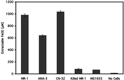
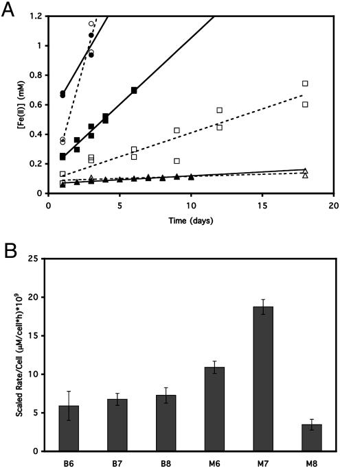
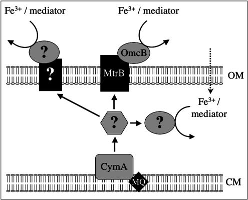
We developed a new method to measure iron reduction at a distance based on depositing Fe(III) (hydr)oxide within nanoporous glass beads. In this "Fe-bead" system, Shewanella oneidensis reduces at least 86.5% of the iron in the absence of direct contact. Biofilm formation accompanies Fe-bead reduction and is observable both macro- and microscopically. Fe-bead reduction is catalyzed by live cells adapted to anaerobic conditions, and maximal reduction rates require sustained protein synthesis. The amount of reactive ferric iron in the Fe-bead system is available in excess such that the rate of Fe-bead reduction is directly proportional to cell density; i.e., it is diffusion limited. Addition of either lysates prepared from anaerobic cells or exogenous electron shuttles stimulates Fe-bead reduction by S. oneidensis, but iron chelators or additional Fe(II) do not. Neither dissolved Fe(III) nor electron shuttling activity was detected in culture supernatants, implying that the mediator is retained within the biofilm matrix. Strains with mutations in omcB or mtrB show about 50% of the wild-type levels of reduction, while a cymA mutant shows less than 20% of the wild-type levels of reduction and a menF mutant shows insignificant reduction. The Fe-bead reduction defect of the menF mutant can be restored by addition of menaquinone, but menaquinone itself cannot stimulate Fe-bead reduction. Because the menF gene encodes the first committed step of menaquinone biosynthesis, no intermediates of the menaquinone biosynthetic pathway are used as diffusible mediators by this organism to promote iron reduction at a distance. CymA and menaquinone are required for both direct and indirect mineral reduction, whereas MtrB and OmcB contribute to but are not absolutely required for iron reduction at a distance.


Figures

FIG. 1.
FIG. 1.
Fe-bead characterization. (A and B) SEM (A) and TEM (B) images of Fe-beads, showing the surface structure, 50-nm-diameter pores, and the size of the pores relative to an S. oneidensis MR-1 cell (on the right side in panel B). (C) Schematic diagram providing the dimensions of the bead core and cortex. (D) Percentages of iron in the core of the beads on days 0 and 3 as determined by TEM and EDS, prior to acid extraction. The error bars indicate the standard deviations for 50 EDS measurements from a single experiment (cell density, 6.7 × 10⁸ cell/ml).
FIG. 2.
FIG. 2.
Reduction of Fe-beads by Shewanella strains. The strains used were S. oneidensis MR-1, Shewanella sp. strain ANA-3, S. putrefaciens CN-32, and E. coli MG1655. Killed MR-1 cells were killed by formaldehyde treatment as described in Materials and Methods. The data are the averages of duplicate experiments, and the error bars indicate the data ranges for 3-day incubations with Fe-beads.
FIG. 3.
FIG. 3.
Biofilm formation on Fe-beads by S. oneidensis MR-1. (A) Inverted tubes showing clumping of inoculated Fe-beads after 24 h of incubation (left tube) and an uninoculated control (right tube). (B) Confocal microscopy image of an MR-1 microcolony on an Fe-bead surface after 3 days. Scale bar = 10 μm. The arrow indicates a microcolony. (C) Environmental scanning electron microscopy images of MR-1 biofilms on the Fe-bead surface. (Top panel) Fe-bead surface in the absence of bacteria; (middle panel) Fe-bead surface after 3 days of incubation with MR-1 cells (scale bar = 20 μm); (bottom panel) close-up of Fe-bead surface after 3 days of incubation with MR-1 cells (scale bar = 2 μm).
FIG. 4.
FIG. 4.
(A) Reduction of Fe-beads by S. oneidensis MR-1 as a function of the preincubation conditions. MR-1 cells were preincubated with 1 mM fumarate (diamonds) or 1 mM ferric citrate (squares) or under oxygen-limited conditions (triangles). (B) Fe-bead reduction by anaerobically and aerobically grown MR-1 cells. Fumarate-grown cells were grown anaerobically with 10 mM fumarate as the electron acceptor, while aerobically grown cells were grown with oxygen as the electron acceptor. Chloramphenicol was added to separate Fe-bead cultures with these cells (+ Cm) to inhibit new protein synthesis. (C) Reduction of Fe-beads as a function of cell density. Serial dilutions of a standard high-density Fe-bead culture inoculum were incubated in separate tubes with Fe-beads. A linear fit to the data is shown. Each data point is the average for duplicate cultures (the error bars indicate the ranges) from 3 days of incubation with the Fe-beads.
FIG. 5.
FIG. 5.
Fe-bead reduction by S. oneidensis MR-1 compared with Fe(III) (hydr)oxide reduction. (A) Time course of iron reduction by 6.7 × 10⁸ (circles), 6.7 × 10⁷ (squares), and 6.7 × 10⁶ (triangles) S. oneidensis MR-1 cells/ml with Fe-beads (open symbols and dashed lines) or free Fe(III) (hydr)oxide particles (solid symbols and solid lines) as the electron acceptor. Each data point represents a separate tube sacrificed at a given time for the measurement. Regression lines were calculated by the least-squares method. The initial amount of iron reduced (first day) was not included in the regressions because we could not exclude direct contact reduction of the iron in the bead cortex. (B) Plot of rates per cell calculated from the regressions shown in panel A for different cell densities with Fe-beads or Fe(III) (hydr)oxide. B6, 6.7 × 10⁶ cells/ml with Fe-beads; B7, 6.7 × 10⁷ cells/ml with Fe-beads; B8, 6.7 × 10⁸ cells/ml with Fe-beads; M6, Fe(III) (hydr)oxide with 6.7 × 10⁶ cells/ml; M7, Fe(III) (hydr)oxide with 6.7 × 10⁷ cells/ml; M8, Fe(III) (hydr)oxide with 6.7 × 10⁸ cells/ml. The error bars indicate the standard errors of the slopes of the regression lines.
FIG. 6.
FIG. 6.
Reduction of Fe-beads by S. oneidensis MR-1 mutants defective in iron reduction. The strains used were strain MR-1 (Wild Type), DKN247 (omcB), DKN248 (mtrB), DKN249 (menF), and DKN250 (cymA). menF + MK-4, strain DKN249 with 10 μM MK-4 added to the anaerobic preincubation with fumarate; menF + DHNA, strain DKN249 with 10 μM DHNA added to the anaerobic preincubation with fumarate. The data indicate the amounts of Fe reduced following our standard 3-day incubation with 6.7 × 10⁸ cells/ml.
FIG. 7.
FIG. 7.
Reduction of Fe(III) (hydr)oxide, ferric citrate, or AQDS by S. oneidensis MR-1 mutants defective in electron transfer. (A) Reduction of 0.5 mM Fe(III) (hydr)oxide; (B) reduction of 1 mM ferric citrate; (C) reduction of 1 mM AQDS. Symbols: ▪, wild-type strain MR-1; ▾, omcB mutant DKN247; ▴, mtrB mutant DKN248; •, menF mutant DKN249; ⋄, cymA mutant DKN250. The error bars indicate the ranges for duplicate cultures for the iron reduction experiments and the standard deviations for triplicate cultures for the AQDS reduction experiment.
FIG. 8.
FIG. 8.
Model for iron reduction at a distance in S. oneidensis MR-1. Chelated Fe(III) or a redox-active mediator is reduced extracellularly by the OmcB-MtrB complex necessary for iron mineral reduction. In the absence of or in addition to this reduction pathway, chelated Fe(III) or the redox-active mediator is reduced by an alternate pathway that may involve other redox-active outer membrane complexes (such as the mtrDEF complex or the putative outer membrane molybdopterin oxidoreductase complexes encoded by the SO1427-SO1432 and SO4362-SO4357 gene clusters). Alternatively, chelated Fe(III) or the redox-active mediator may be transported into the bacterial periplasm and reduced by periplasmic electron transport components, such as low-potential multiheme c-type cytochromes. OM, outer membrane; MQ, menaquinone; CM, cytoplasmic membrane.

References

    1. Assfalg, M., I. Bertini, M. Bruschi, C. Michel, and P. Turano. 2002. The metal reductase activity of some multiheme cytochromes c: NMR structural characterization of the reduction of chromium(VI) to chromium(III) by cytochrome c7. Proc. Natl. Acad. Sci. USA 99:9750-9754. - PMC - PubMed
    1. Beliaev, A. S., and D. A. Saffarini. 1998. Shewanella putrefaciens mtrB encodes an outer membrane protein required for Fe(III) and Mn(IV) reduction. J. Bacteriol. 180:6292-6297. - PMC - PubMed
    1. Beliaev, A. S., D. A. Saffarini, J. L. McLaughlin, and D. Hunnicutt. 2001. MtrC, an outer membrane decahaem c cytochrome required for metal reduction in Shewanella putrefaciens MR-1. Mol. Microbiol. 39:722-730. - PubMed
    1. Beveridge, T. J., S. A. Makin, J. L. Kadurugamuwa, and Z. S. Li. 1997. Interactions between biofilms and the environment. FEMS Microbiol. Rev. 20:291-303. - PubMed
    1. Blattner, F. R., G. Plunkett, C. A. Bloch, N. T. Perna, V. Burland, M. Riley, J. ColladoVides, J. D. Glasner, C. K. Rode, G. F. Mayhew, J. Gregor, N. W. Davis, H. A. Kirkpatrick, M. A. Goeden, D. J. Rose, B. Mau, and Y. Shao. 1997. The complete genome sequence of Escherichia coli K-12. Science 277:1453-1474. - PubMed

Publication types