Biodesulfurization of dibenzothiophene by microbial cells coated with magnetite nanoparticles
- PMID: 16085841
- PMCID: PMC1183266
- DOI: 10.1128/AEM.71.8.4497-4502.2005
Biodesulfurization of dibenzothiophene by microbial cells coated with magnetite nanoparticles
Abstract
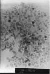
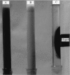
Microbial cells of Pseudomonas delafieldii were coated with magnetic Fe3O4 nanoparticles and then immobilized by external application of a magnetic field. Magnetic Fe3O4 nanoparticles were synthesized by a coprecipitation method followed by modification with ammonium oleate. The surface-modified Fe3O4 nanoparticles were monodispersed in an aqueous solution and did not precipitate in over 18 months. Using transmission electron microscopy (TEM), the average size of the magnetic particles was found to be in the range from 10 to 15 nm. TEM cross section analysis of the cells showed further that the Fe3O4 nanoparticles were for the most part strongly absorbed by the surfaces of the cells and coated the cells. The coated cells had distinct superparamagnetic properties. The magnetization (delta(s)) was 8.39 emu.g(-1). The coated cells not only had the same desulfurizing activity as free cells but could also be reused more than five times. Compared to cells immobilized on Celite, the cells coated with Fe3O4 nanoparticles had greater desulfurizing activity and operational stability.
Figures


Similar articles
-
In situ magnetic separation and immobilization of dibenzothiophene-desulfurizing bacteria.Bioresour Technol. 2009 Nov;100(21):5092-6. doi: 10.1016/j.biortech.2009.05.064. Epub 2009 Jun 21. Bioresour Technol. 2009. PMID: 19541480
-
Biodesulfurization using Pseudomonas delafieldii in magnetic polyvinyl alcohol beads.Lett Appl Microbiol. 2005;40(1):30-6. doi: 10.1111/j.1472-765X.2004.01617.x. Lett Appl Microbiol. 2005. PMID: 15612999
-
Optimal design and characterization of superparamagnetic iron oxide nanoparticles coated with polyvinyl alcohol for targeted delivery and imaging.J Phys Chem B. 2008 Nov 20;112(46):14470-81. doi: 10.1021/jp803016n. Epub 2008 Aug 26. J Phys Chem B. 2008. PMID: 18729404
-
Magnetic immobilization of bacteria using iron oxide nanoparticles.Biotechnol Lett. 2018 Feb;40(2):237-248. doi: 10.1007/s10529-017-2477-0. Epub 2017 Nov 27. Biotechnol Lett. 2018. PMID: 29181762 Review.
-
[Frontier of chemical modification of protein: application of polyethyleneglycol-modified enzymes in biotechnological processes].Seikagaku. 1988 Sep;60(9):1005-18. Seikagaku. 1988. PMID: 3073171 Review. Japanese. No abstract available.
Cited by
-
The surfactant tween 80 enhances biodesulfurization.Appl Environ Microbiol. 2006 Nov;72(11):7390-3. doi: 10.1128/AEM.01474-06. Epub 2006 Sep 15. Appl Environ Microbiol. 2006. PMID: 16980422 Free PMC article.
-
Effects of Modified Magnetite Nanoparticles on Bacterial Cells and Enzyme Reactions.Nanomaterials (Basel). 2020 Jul 30;10(8):1499. doi: 10.3390/nano10081499. Nanomaterials (Basel). 2020. PMID: 32751621 Free PMC article.
-
Improvement of biodesulfurization activity of alginate immobilized cells in biphasic systems.J Ind Microbiol Biotechnol. 2008 Mar;35(3):145-50. doi: 10.1007/s10295-007-0268-7. Epub 2007 Nov 6. J Ind Microbiol Biotechnol. 2008. PMID: 17985163
-
Biofilm inhibition, modulation of virulence and motility properties by FeOOH nanoparticle in Pseudomonas aeruginosa.Braz J Microbiol. 2019 Jul;50(3):791-805. doi: 10.1007/s42770-019-00108-z. Epub 2019 Jun 27. Braz J Microbiol. 2019. PMID: 31250405 Free PMC article.
-
Mechanistic and recent updates in nano-bioremediation for developing green technology to alleviate agricultural contaminants.Int J Environ Sci Technol (Tehran). 2022 Sep 29:1-26. doi: 10.1007/s13762-022-04560-7. Online ahead of print. Int J Environ Sci Technol (Tehran). 2022. PMID: 36196301 Free PMC article. Review.
References
-
- Amanda, K. Y., and K. D. Wisecarver. 1992. Cell immobilization using PVA crosslinked with boric acid. Biotechnol. Bioeng. 39:447-449. - PubMed
-
- Beverly, L. M. F. 1999. Biodesulfurization. Curr. Opin. Microbiol. 2:257-264. - PubMed
-
- Býlkova, Z., M. Slovakova, D. Horak, J. Lenfeld, and J. Churácek. 2002. Enzymes immobilized on magnetic supports: efficient and selective system for protein modification. J. Chromatogr. B 770:177-181. - PubMed
-
- Chang, J. H., Y. K. Chang, H. W. Ryu, and H. N. Chang. 2000. Desulfurization of light gas oil in immobilized-cell systems of Gordona sp. CYKS1 and Nocardia sp. CYKS2. FEMS Microbiol. Lett. 182:309-312. - PubMed
-
- Chotani, G. K., and A. Constaninides. 1984. Immobilized cell cross-flow reactor. Biotechnol. Bioeng. 26:217-220. - PubMed
Publication types
MeSH terms
Substances
LinkOut - more resources
Full Text Sources
Other Literature Sources
Medical

