. 2005 Aug;12(8):959-69.
doi: 10.1128/CDLI.12.8.959-969.2005.

Optimization and validation of a multiplexed luminex assay to quantify antibodies to neutralizing epitopes on human papillomaviruses 6, 11, 16, and 18

Affiliations

Optimization and validation of a multiplexed luminex assay to quantify antibodies to neutralizing epitopes on human papillomaviruses 6, 11, 16, and 18

Dennis Dias et al. Clin Diagn Lab Immunol. 2005 Aug.

Abstract

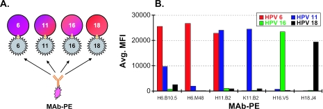
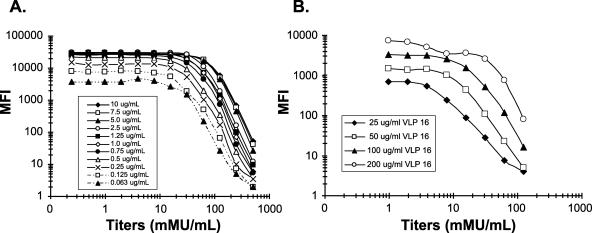
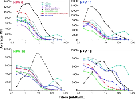
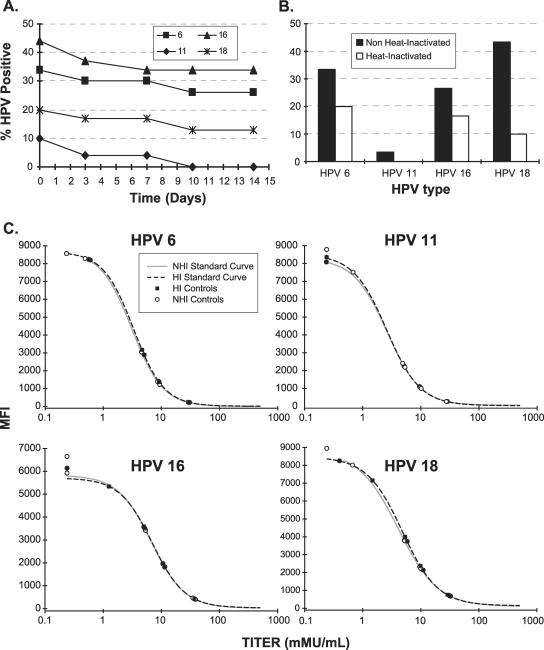
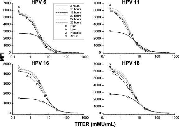
A human papillomavirus (HPV) multiplexed competitive Luminex immunoassay first described by Opalka et al. (D. Opalka, C. E. Lachman, S. A. MacMullen, K. U. Jansen, J. F. Smith, N. Chirmule, and M. T. Esser, Clin. Diagn. Lab. Immunol. 10:108--15, 2003) was optimized and validated for use in epidemiology studies and vaccine clinical trials. Optimization increased both the analytical sensitivity and the clinical specificity of the assay to more effectively discriminate the low-titer antibody response of HPV-infected persons from noninfected individuals. The characteristics of the assay that were optimized included monoclonal antibody (MAb) specificity, scaling up the conjugation of virus-like particles (VLPs) to microspheres, VLP concentration, MAb concentration, sample matrix, sample dilution, incubation time, heat inactivation of sample sera, and detergent effects on assay buffer. The assay was automated by use of a TECAN Genesis Workstation, thus improving assay throughput, reproducibility, and operator safety. Following optimization, the assay was validated using several distinct serum panels from individuals determined to be at low and high risk for HPV infection. The validated assay was then used to determine the clinical serostatus cutoff. This high-throughput assay has proven useful for performing epidemiology studies and evaluating the efficacy of prophylactic HPV vaccines.


Figures

FIG. 1.
FIG. 1.
Monoclonal antibody specificity. (A) Experimental design for MAb specificity experiment. A single MAb-PE was incubated with VLP 6, 11, 16, and 18 microspheres. Detection of MAb-PE to any of the four VLP types revealed any cross-reactivity. (B) MAb specificity results. HPV 6, 11, 16, and 18 VLP-microspheres (5,000 VLP-microspheres per type; VLP concentration, 200 μg/ml) were added in quadriplex to each well. One MAb-PE was added to each well at a concentration of 40 μg/ml. Detection of fluorescence from the PE-labeled MAbs is shown in median fluorescence intensity (MFI).
FIG. 2.
FIG. 2.
Effect of VLP and monoclonal antibody-PE concentration on assay sensitivity for HPV 16. (A) Effect of different concentrations of H16.V5 MAb-PE (0.0625 to 10.0 μg/ml) on assay sensitivity and dynamic range. VLP-microspheres used in this experiment were coupled with HPV 16 VLP at a concentration of 200 μg/ml. (B) Effect of different concentrations of VLP 16 (25 to 200 μg/ml) on assay sensitivity and dynamic range. The concentration of H16.V5 MAb-PE was 0.1 μg/ml.
FIG. 3.
FIG. 3.
Effect of different sample matrices on assay sensitivity and robustness. Each standard curve was generated using the specified matrix, 100 μg/ml VLP-microspheres, and a 0.1-μg/ml final MAb-PE concentration. The mixture was incubated overnight (15 to 25 h) and then qualitatively and quantitatively evaluated in comparison to normal human serum (NHS). Abbreviations: bovine serum albumin (BSA), recombinant human albumin (rHA), normal human serum (NHS), normal goat serum (NGS), antibody depleted human serum (ADHS), and defibrinated plasma (DFP).
FIG. 4.
FIG. 4.
Effect of heat inactivation of serum samples on HPV antibody titers. (A) Seventy randomly selected retention samples that had never been thawed were thawed at RT and stored at 4°C. The samples were tested in the cLIA on days 0, 3, 7, 10, and 14. (B) Percentage of HPV-positive samples using a serostatus cutoff of 24 mMU/ml for all HPV types. A total of 70 freshly thawed samples were aliquoted into two tubes. One of the tubes was heat inactivated for 30 ± 2 min at 56°C, while the other remained at 4°C. Both samples were tested on the same plate in duplicate and read from the same standard curve. (C) Heat inactivation of reference serum has no effect on antibody titers. Reference serum was either heat inactivated or remained at 4°C before spiking into ADHS and tested in the HPV cLIA.
FIG. 5.
FIG. 5.
Effect of different assay incubation times on assay sensitivity, dynamic range, precision, and robustness. Each curve on the graph represents a four-parameter (4-PL) logistic fit of a 12-point standard curve for HPV 6, 11, 16, and 18. Antibody titers generated from the controls of each standard curve have been plotted on top of each standard curve. VLP-microspheres used in this experiment were coupled with 100 μg/ml of HPV 16 VLP and 25 μg/ml of HPV 6, 11, and 18 VLPs. The concentrations of H6.M48-PE, K11.B2-PE, H16.V5-PE, and H18.J4-PE were 0.1 μg/ml. All standards were at a final starting concentration of 500 mMU/ml. The mixture of VLP-microspheres, MAb-PE, reference sera, and ADHS was incubated for 3, 15, 18, 20, 22, or 25 h.

References

    1. American Cancer Society. 2004. Cancer facts and figures 4. American Cancer Society, Atlanta, Ga.
    1. Atypical Squamous Cells of Undetermined Significance/Low-Grade Squamous Intraepithelial Lesions Triage Study (ALTS) Group. 2000. Human papillomavirus testing for triage of women with cytologic evidence of low-grade squamous intraepithelial lesions: baseline data from a randomized trial. J. Natl. Cancer Inst. 92:397-402. - PubMed
    1. Bosch, F. X., A. Lorincz, N. Munoz, C. J. Meijer, and K. V. Shah. 2002. The causal relation between human papillomavirus and cervical cancer. J. Clin. Pathol. 55:244-265. - PMC - PubMed
    1. Bosch, F. X., M. M. Manos, N. Munoz, M. Sherman, A. M. Jansen, J. Peto, M. H. Schiffman, V. Moreno, R. Kurman, and K. V. Shah. 1995. Prevalence of human papillomavirus in cervical cancer: a worldwide perspective. International biological study on cervical cancer (IBSCC) study group. J. Natl. Cancer Inst. 87:796-802. - PubMed
    1. Carter, J. J., L. A. Koutsky, J. P. Hughes, S. K. Lee, J. Kuypers, N. Kiviat, and D. A. Galloway. 2000. Comparison of human papillomavirus types 16, 18, and 6 capsid antibody responses following incident infection. J. Infect. Dis. 181:1911-1919. - PubMed

Publication types

MeSH terms

LinkOut - more resources