. 2005 Nov 15;392(Pt 1):231-9.
doi: 10.1042/BJ20050578.

Human T-cell lymphotropic virus type I-transformed T-cells have a partial defect in ceramide synthesis in response to N-(4-hydroxyphenyl)retinamide

Affiliations

Human T-cell lymphotropic virus type I-transformed T-cells have a partial defect in ceramide synthesis in response to N-(4-hydroxyphenyl)retinamide

Nadine Darwiche et al. Biochem J. .

Erratum in

  • Biochem J. 2012 Jun 15;444(3):619. Abou Tayyoun, Ahmad [corrected to Tayoun, Ahmad Abou]

Abstract

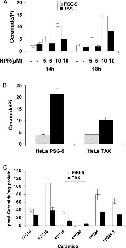
Treatment with the synthetic retinoid HPR [N-(4-hydroxyphenyl)-retinamide] causes growth arrest and apoptosis in HTLV-I (human T-cell lymphotropic virus type-I)-positive and HTLV-I-negative malignant T-cells. It was observed that HPR-mediated growth inhibition was associated with ceramide accumulation only in HTLV-I-negative cells. The aim of the present study was to investigate the mechanism by which HPR differentially regulates ceramide metabolism in HTLV-I-negative and HTLV-I-positive malignant T-cells. Clinically achievable concentrations of HPR caused early dose-dependent increases in ceramide levels only in HTLV-I-negative cells and preceded HPR-induced growth suppression. HPR induced de novo synthesis of ceramide in HTLV-I-negative, but not in HTLV-I-positive, cells. Blocking ceramide glucosylation in HTLV-I-positive cells, which leads to accumulation of endogenous ceramide, rendered these cells more sensitive to HPR. Exogenous cell-permeant ceramides that function partially by generating endogenous ceramide induced growth suppression in all tested malignant lymphocytes, were consistently found to be less effective in HTLV-I-positive cells confirming their defect in de novo ceramide synthesis. Owing to its multipotent activities, the HTLV-I-encoded Tax protein was suspected to inhibit ceramide synthesis. Tax-transfected Molt-4 and HELA cells were less sensitive to HPR and C6-ceramide mediated growth inhibition respectively and produced lower levels of endogenous ceramide. Together, these results indicate that HTLV-I-positive cells are defective in de novo synthesis of ceramide and that therapeutic modalities that bypass this defect are more likely to be successful.


Figures

Figure 1
Figure 1. HPR induces distinct ceramide responses in HTLV-I-negative and HTLV-I-positive malignant human T-cell lines
(A) Ceramide levels in the HTLV-I-negative CEM human T-cell line in response to HPR. CEM cells were seeded at a density of 3.5×10⁵ cells/ml and treated with 0.1% DMSO as a control or with 1 μM HPR for the times indicated. Ceramide levels were determined using the DGK assay as described in the Experimental section and normalized to total cellular lipid phosphate levels. Data points represent the mean±range (n=2). Results are representative of three independent experiments. (B) Effects of HPR treatment on the growth of CEM human T-cell line. Cells were seeded at a density of 2×10⁵ cells/ml and treated with 0.1% DMSO as a control or with 1 μM HPR for the times indicated. Viable cell counts, calculated from triplicate wells by Trypan Blue dye exclusion, are expressed as a percentage of controls and are representative of two independent experiments. (C) Dose–response to HPR treatment in CEM human T-cell line. Cells were seeded at a density of 3.5×10⁵ cells/ml and treated with 0.1% DMSO as a control, or the indicated concentrations of HPR, for 24 h. Ceramide levels were determined as in (A). Data points represent the mean±range (n=2). Results are representative of three independent experiments. (D) HPR treatment induces late ceramide accumulation in HTLV-I-positive human T-cell line Hut-102. Cells were seeded at a density of 3.5×10⁵ cells/ml and treated with 0.1% DMSO as a control or 5 μM HPR for the times indicated. Ceramide levels were determined as in (A). Data points represent the mean±range (n=2). Results are representative of two independent experiments. (E) Dose–response to HPR treatment in HTLV-I-positive human T-cell line C8166. Cells were seeded at a density of 3.5×10⁵ cells/ml and treated with the indicated concentrations of HPR for 48 h. Ceramide levels were determined as in (A). Data points represent the mean±range (n=2). Results are representative of two independent experiments.
Figure 2
Figure 2. HPR treatment induces de novo synthesis of ceramide in HTLV-I-negative human T-cell lines only
CEM and C8166 cells were seeded at a density of 3.5×10⁵ cells/ml and treated with 0.1% DMSO as a control or with 1 and 5 μM HPR respectively. De novo ceramide levels were determined using the [3H]palmitate incorporation method as described in the Experimental section and normalized to total cellular lipid phosphate levels. Data points represent the mean±range (n=2) and are percentages of treated over control cells. Results are representative of two independent experiments.
Figure 3
Figure 3. PDMP synergizes with HPR treatment to induce cell death in HTLV-I-positive human T-cell lines
(A) Ceramide levels in the HTLV-I-positive C8166 and HuT-102 cell lines in response to combined HPR and PDMP treatment. Cells were seeded at a density of 3.5×10⁵ cells/ml and were pretreated with 20 μM PDMP, an inhibitor of GCS. PDMP pretreatment was performed 2 h before treatment with 1 μM HPR. Data points represent the mean±range (n=2). Results are representative of two independent experiments. (B) Effect of combined HPR and PDMP treatment on the growth of the HTLV-I-positive C8166 and HuT-102 cells. Cells were seeded at a density of 2×10⁵ cells/ml and were pretreated with 20 μM PDMP, an inhibitor of GCS. PDMP pretreatment was performed 2 h prior to treatment with 1 μM HPR. Viability was assayed in 96-well plates at 24 h using the Cell Titer 96® non-radioactive cell proliferation kit. Data points represent the mean±S.D. (n=3). Results are expressed as percentage of control cells (0.1% DMSO) and represent the mean of three independent experiments.
Figure 4
Figure 4. Response to exogenous C6-ceramide in HTLV-I-positive human T-cell lines is blunted because of a defect in endogenous ceramide synthesis
(A) Endogenous ceramide levels in the HTLV-I-positive (HuT-102, MT2 and C8166) and the HTLV-I-negative (CEM, Jurkat and Molt-4) human T-cell lines in response to C6-ceramide. Cells were seeded in RPMI-1640 medium containing 2% FBS at a density of 3.5×10⁵ cell/ml. Cells were treated with 20 μM C6-ceramide for 18 h. Endogenous ceramide levels were determined as in Figure 1(A). The phosphorylated product of C6-ceramide is easily distinguishable from that of endogenous ceramide on the TLC plate (results not shown). Data points represent the mean±range (n=2). Results are representative of two independent experiments. (B) Effects of C6-ceramide on the growth of the HTLV-I-positive (HuT-102 and MT2) and the HTLV-I-negative (CEM, Jurkat and Molt-4) human T-cell lines. Cells were seeded in RPMI-1640 medium containing 2% FBS at a density of 2×10⁵ cells/ml. Cells were treated with 20 μM C6-ceramide for 18 h. Viability was assayed using the Trypan Blue dye exclusion method. Data points represent the mean±S.D. (n=3). Results are expressed as percentage of control cells (0.1% ethanol) and represent the mean of two independent experiments. (C) Endogenous ceramide levels in the HTLV-I-positive HuT-102 and the HTLV-I-negative Jurkat human T-cell lines in response to C6-ceramide and FB1 treatment. Cells were seeded in RPMI-1640 medium containing 2% FBS at a density of 3.5×10⁵ cell/ml. Cells were treated with 20 μM C6-ceramide, 50 μM FB1, or combined C6-ceramide and FB1 for 18 h. Endogenous ceramide levels were determined as in Figure 1(A). Data points represent the mean±range (n=2). Results are representative of two independent experiments. (D) Endogenous ceramide levels in the HTLV-I-positive HuT-102 and the HTLV-I-negative Jurkat human T-cell lines in response to exogenous C2-ceramide. Cells were seeded in RPMI-1640 medium containing 2% FBS at a density of 3.5×10⁵ cell/ml. Cells were treated with 20 μM C2-ceramide for 18 h. Endogenous ceramide levels were determined as in Figure 1(A). Data points represent the mean±range (n=2). Results are representative of two independent experiments.
Figure 5
Figure 5. Role of Tax protein in suppressing the ceramide response in the HTLV-I-positive human T-cell lines
(A) Reduced ceramide levels in HPR-treated Tax-transfected Molt-4 cells. Molt-4 cells were transiently transfected with pSG5-Tax or control vector and treated after 24 h with HPR (5 μM and 10 μM) for the times indicated. Ceramide levels were determined as in Figure 1(A). Data points represent the mean±range (n=2). Results are representative of two independent experiments. (B) Effects of exogenous C6-ceramide on endogenous ceramide levels in Tax-transfected HeLa cells. pSG5-Tax or control cells were transfected for 24 h and then were treated with 20 μM C6-ceramide in DMEM medium containing 2% FBS for 18 h. Endogenous ceramide levels were determined as in Figure 1(A). Data points represent the mean±range (n=2). Results are representative of two independent experiments. (C) Ceramide synthase activity is reduced in Tax-transfected Hela cells. pSG5-Tax or control cells were transfected for 24 h and then were treated with 5 μM 17C6-ceramide in DMEM medium containing 2% FBS for 24 h. Endogenous 17-ceramide species levels were determined as described in the Experimental section and normalized to total cellular protein levels. Data points represent the mean±range (n=2).

Similar articles

Cited by

References

    1. Delia D., Aiello A., Lombardi L., Pelicci P. G., Grignani F., Grignani F., Formelli F., Menard S., Costa A., Veronesi U. N-(4-hydroxyphenyl)retinamide induces apoptosis of malignant hemopoietic cell lines including those unresponsive to retinoic acid. Cancer Res. 1993;53:6036–6041. - PubMed
    1. Wu J. M., DiPietrantonio A. M., Hsieh T. C. Mechanism of fenretinide (4-HPR)-induced cell death. Apoptosis. 2001;6:377–388. - PubMed
    1. Kalemkerian G. P., Slusher R., Ramalingam S., Gadgeel S., Mabry M. Growth inhibition and induction of apoptosis by fenretinide in small-cell lung cancer cell lines. J. Natl. Cancer Inst. 1995;87:1674–1680. - PubMed
    1. Maurer B. J., Metelitsa L. S., Seeger R. C., Cabot M. C., Reynolds C. P. Increase of ceramide and induction of mixed apoptosis/necrosis by N-(4-hydroxyphenyl)-retinamide in neuroblastoma cell lines. J. Natl. Cancer Inst. 1999;91:1138–1146. - PubMed
    1. Oridate N., Suzuki S., Higuchi M., Mitchell M. F., Hong W. K., Lotan R. Involvement of reactive oxygen species in N-(4-hydroxyphenyl)retinamide-induced apoptosis in cervical carcinoma cells. J. Natl. Cancer Inst. 1997;89:1191–1198. - PubMed

Publication types

MeSH terms