Review
. 2005 May 29;360(1457):903-11.
doi: 10.1098/rstb.2005.1640.

Relating connectional architecture to grey matter function using diffusion imaging

Affiliations
Review

Relating connectional architecture to grey matter function using diffusion imaging

T E J Behrens et al. Philos Trans R Soc Lond B Biol Sci. .

Abstract

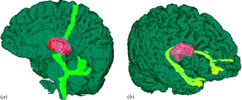
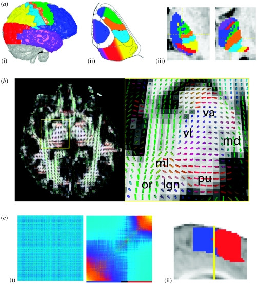
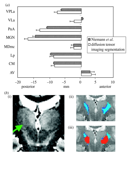
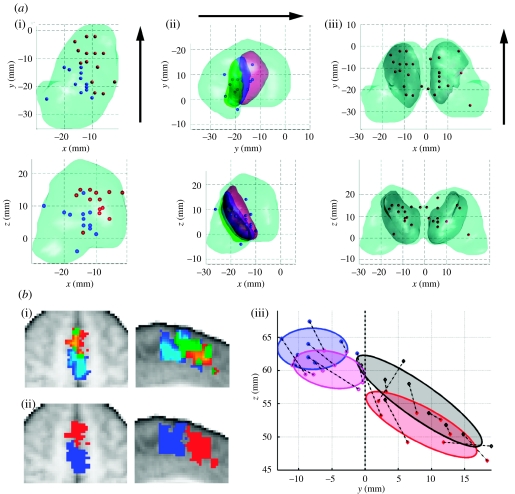
Understanding brain function in terms of connectional architecture is a major goal of neuroimaging. However, direct investigation of the influence of brain circuitry on function has been hindered by the lack of a technique for exploring anatomical connectivity in the in vivo brain. Recent advances in magnetic resonance diffusion imaging have given scientists access to data relating to local white matter architecture and, for the first time, have raised the possibility of in vivo investigations into brain circuitry. This review investigates whether diffusion imaging may be used to identify regions of grey matter that are distinct in their connectional architecture, and whether these connectional differences are reflected either in local cytoarchitecture or in local grey matter function. Establishing a direct relationship between regional boundaries based on diffusion imaging and borders between regions that perform different functions would not only be of great significance when interpreting functional results, but would also provide a first step towards the validation of diffusion-based anatomical connectivity studies.


Figures

Figure 1
Figure 1
Tracing anatomical connections with diffusion imaging: (a) seeded from a single voxel in a ventral lateral location in thalamus. In macaque monkeys, ventral lateral nucleus of thalamus (VL) processes motor information and projects to primary motor cortex (M1; Jones 1985). The connectivity distribution both ascended to the anterior bank of the central sulcus (M1) and descended. The descending distribution followed two distinct paths, one entered the cerebellum and branched, terminating in the cerebellar cortex, the other continued further down the brainstem. (b) Seeded from a single voxel in a medial dorsal location in thalamus. In macaque monkeys, mediodorsal nucleus of thalamus is associated with cognitive processing, and is reciprocally connected to prefrontal (PFC; Tanaka 1976) and temporal cortices (Markowitsch et al. 1985). The connectivity distribution progressed anteriorly to the lateral prefrontal cortex and also, at first posteriorly, around the posterior edge of the thalamus, and then anteriorly to the anterior temporal cortex. In each case, the thalamic seed voxel is strongly connected to cortical regions relevant to its function. The two seed voxels, which are functionally distinct, may also be distinguished on the basis of their connectivity distributions. Based on Behrens et al. (2003a).
Figure 2
Figure 2
Diffusion-based parcellation of grey matter: (a) Identifying thalamic nuclei on the basis of their remote cortical projections. (i) Cortex was first segmented into large anatomically defined regions corresponding to known connection areas of the major thalamic nuclear groups in non-human primates. Subsequently, probabilistic tractography was seeded from each thalamic voxel, and the probability of connection from that voxel to each of the cortical regions was recorded. (ii) Shows an axial section based on a histological atlas of the human thalamus with nuclei outlined by black lines (Morel et al. 1997). Nuclei have been colour-coded according to the cortical zone with which they are expected to show the strongest connections on the basis of the non-human literature. (iii) Shows axial (left) and coronal (right) sections through a human thalamus in a single subject, with each thalamic voxel colour-coded according to the cortical zone with which it had the highest probability of connection. (figure based on Behrens et al. 2003a). (b) Identifying thalamic nuclei on the basis of their local diffusion properties. Left: cuboid rendering of diffusion tensors in a single slice through a human brain. Cuboids are coloured according to the orientation of their principal axis. Red corresponds to medial–lateral; green to anterior–posterior; blue to superior–inferior. Right: close-up through thalamus. Clusters are identified according to the diffusion tensor in each voxel. Nuclei labels were assigned based on the location and fibre orientation of the clusters (Wiegell et al. 2003). The lateral geniculate nucleus (lgn; purple) bends though Meyer's loop (ml; purple turning green) to the optic radiation (or; green). The pulvinar nucleus (pu; red) projects medial-laterally and the mediodorsal nucleus (md; green) anteriorly. The ventral lateral nucleus (vl; blue-purple) projects superior-laterally and the ventral anterior nucleus (va) superiorly. Courtesy of Ulas Ziyan (MIT) and David Tuch (Harvard Medical School and Massachusetts General Hospital). (c) Identifying the boundary between SMA and preSMA on the basis of a sharp change in connectivity profile. Connectivity profiles are seeded from every voxel in an area including SMA and preSMA. The correlation between each pair of profiles is stored in a matrix ((i) left) whose nodes are reordered according to a spectral reordering routine (Johansen-Berg et al. 2004). Groups of commonly connected clusters are clearly visible in the reordered matrix ((i) right) and are identified by the colour bar below the matrix. These clusters map back to spatially contiguous regions of cortex aligned along the anterior–posterior axis. The division between the clusters is close to the vertical line from the anterior commisure (y=0 mm in MNI space; Evans et al. 1992—yellow line in (ii))—the best approximation for the division between human SMA and preSMA (Zilles et al. 1996). (Figure based on Johansen-Berg et al. (2004), © 2004 the National Academy of Sciences.)
Figure 3
Figure 3
Structural validation of diffusion-based grey matter parcellations: (a; taken from Wiegell et al. 2003) Comparison between centres of gravity of thalamic nuclei defined on the basis of local diffusion characteristics (white bars), and the same nuclei defined histologically on the basis of local fibre orientation (hashed bars; Niemann et al. 2000). Coordinates are along the anterior–posterior axis of the MNI-152 average brain (Evans et al. 1992). Similar results are available for the medial–lateral and superior–inferior axes (Wiegell et al. 2003). (Figure taken from Wiegell et al. (2003).) (b; Sillery et al. in preparation) Comparison between ventral–posterior nucleus of thalamus defined by remote connectivity to sensory cortices, and by contrast enhanced structural MRI (Sillery et al. in preparation). (i) Proton density-weighted MRI shows a hypo-intense region corresponding to the ventral posterior nucleus of thalamus (green arrow). (ii) Region of thalamus defined by 25% chance of connection to sensory cortices. Data from macaque predict that this region will correspond to ventral posterior nucleus. (iii) Manually defined outline of ventral posterior nucleus drawn on (i) and overlaid on (ii). (Figure courtesy of Emma Sillery, FMRIB Centre, University of Oxford.)
Figure 4
Figure 4
Functional relevance of diffusion-based parcellations. (a) Functional relevance of connectivity-based parcellation of thalamus. In each figure, the pale grey surface represents the thalamus; the black arrow represents a posterior–anterior orientation; spheres represent reported centres of functional activation in executive (red) and sensorimotor (blue) tasks; inner semi-transparent surfaces represent thalamic regions in which at least four out of 11 subjects had a probability of 25% of connection to a particular cortical zone. (i) Spatial dissociation between reported thalamic centres of functional activation in sensorimotor and executive tasks. (ii) Thalamic centres of functional activation in sensorimotor tasks aligned well with regions defined by connectivity to sensory (green), primary motor (blue) and premotor (red) cortices. (iii) Thalamic centres of functional activation in executive tasks aligned well with regions defined by connectivity to prefrontal cortex (dark grey). (Figure taken from Johansen-Berg et al. 2005.) (b) Functional relevance of connectional dissociation in medial area 6. (i) Simple motor and cognitive tasks selectively activate SMA (dark to light blue) and preSMA (red to yellow). (ii) Seed voxels which were activated by either cognitive or motor tasks were submitted to connectivity-based parcellation (see figure 2 and main text). A connectional dissociation was found which matched the functional dissociation in (i). (iii) Correspondence between centres of gravity of functionally defined (magenta and black) and connectionally defined (blue and red) SMA and preSMA in nine subjects. Coordinates are in MNI space (Evans et al. 1992) on the mid-sagittal plane. (Figure based on Johansen-Berg et al. (2004), ©2004 by the National Academy of Sciences.)

References

    1. Alexander D.C, Barker G.J, Arridge S.R. Detection and modeling of non-Gaussian apparent diffusion coefficient profiles in human brain data. Magn. Reson. Med. 2002;48:331–340. - PubMed
    1. Amunts K, Schleicher A, Burgel U, Mohlberg H, Uylings H.B, Zilles K. Broca's region revisited: cytoarchitecture and intersubject variability. J. Comp. Neurol. 1999;412:319–341. - PubMed
    1. Basser P, Pierpaoli C. Microstructural and physiological features of tissues elucidated by quantitative-diffusion-tensor MRI. J. Magn. Reson. B. 1996;111:209–219. - PubMed
    1. Basser P.J, Matiello J, Le Bihan D. Estimation of the effective self-diffusion tensor from the NMR spin echo. J. Magn. Reson. B. 1994;103:247–254. - PubMed
    1. Behrens T.E.J, et al. Non-invasive mapping of connections between human thalamus and cortex using diffusion imaging. Nat. Neurosci. 2003a;6:750–757. - PubMed

MeSH terms