. 2005 Oct 14;280(41):34447-57.
doi: 10.1074/jbc.M505229200. Epub 2005 Aug 8.

Identification of the surfactant protein A receptor 210 as the unconventional myosin 18A

Affiliations

Identification of the surfactant protein A receptor 210 as the unconventional myosin 18A

Ching-Hui Yang et al. J Biol Chem. .

Abstract

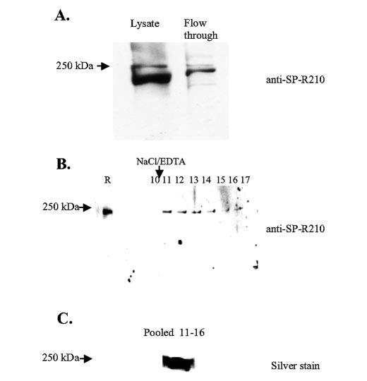
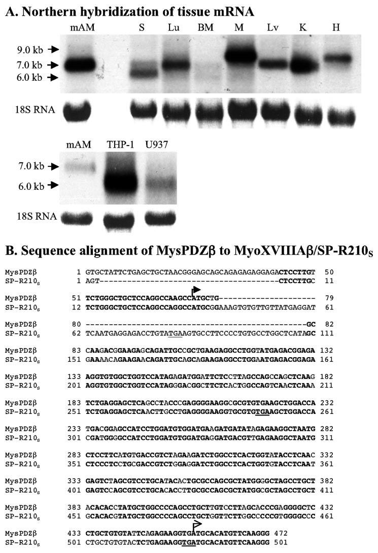
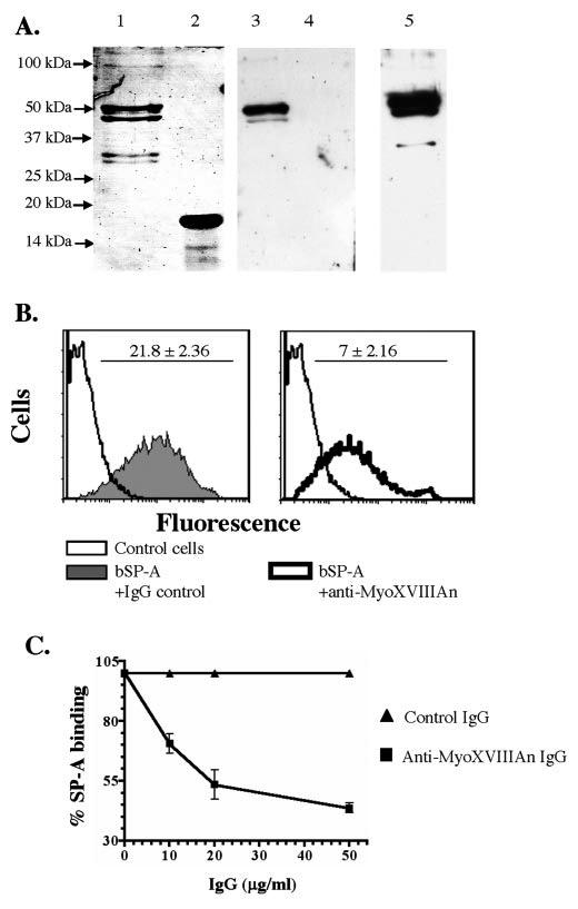
Mass spectrometric characterization of the surfactant protein A (SP-A) receptor 210 (SP-R210) led to the identification of myosin (Myo) XVIIIA and nonmuscle myosin IIA. Antibodies generated against the unique C-terminal tail of MyoXVIIIA revealed that MyoXVIIIA, MyoIIA, and SP-R210 have overlapping tissue distribution, all being highly expressed in myeloid cells, bone marrow, spleen, lymph nodes, and lung. Western blot analysis of COS-1 cells stably transfected with either MyoXVIIIA or MyoIIA indicated that SP-R210 antibodies recognize MyoXVIIIA. Furthermore, MyoXVIIIA but not MyoIIA localized to the surface of COS-1 cells, and most importantly, expression of MyoXVIIIA in COS-1 cells conferred SP-A binding. Western analysis of recombinant MyoXVIIIA domains expressed in bacteria mapped the epitopes of previously derived SP-R210 antibodies to the neck region of MyoXVIIIA. Antibodies raised against the neck domain of MyoXVIIIA blocked the binding of SP-A to macrophages. Together, these findings indicate that MyoXVIIIA constitutes a novel receptor for SP-A.


Figures

FIGURE 1
FIGURE 1
Purification of mouse SP-R210. Mouse SP-R210 was affinity-purified from mAM cells using immobilized biotinylated SP-A as described under “Experimental Procedures.” A, detection of SP-R210 by Western analysis in unprocessed cell lysate and the flow-through eluent after affinity chromatography. B, Western analysis of SP-R210 fractions eluted by 1m NaCl, 10 mmEDTA (lanes 10-17). Lane R indicates SP-R210 retained on the column. C, silver staining of concentrated SP-R210 from the size-fractionated pooled fractions. For Western analysis proteins were fractionated on 8-10% SDS-polyacrylamide gels. Purified SP-R210 was electrophoresed on a 3-8% Tris acetate gel.
FIGURE 2
FIGURE 2
Primary structure analysis of MyoXVIIIA. A, N-terminal 485 amino acids of the longest MyoXVIIIA contain a KE motif, a PDZ domain, clusters of phosphorylation (PO3) sites for casein kinases, protein kinases A and B, and a proline-rich SH3-binding site. An N-terminal KE-rich sequence is present in MysPDZ. Additional phosphorylation sites were also detected in the motor domains and the unique C terminus. The myosin heavy chain motor domain is found between amino acids 485 and 1186. The neck region between amino acids 1186 and 1246 contains an IQ motif and a C-mannosylation site. The dimeric coiled-coil domain spans amino acids 1246 and 1938, and the N-terminal sequence from 1938 to 2035/2054 is unique to MyoXVIIIA. The square box in the C terminus indicates an alternatively spliced coiled-coil insertion. The vertical shaded box in the motor domain outlines a putative transmembrane helix (TM). The short isoform of MyoXVIIIA lacks the N-terminal 485 amino acids. The long and short isoforms are designated as MyoXVIIIAα/MysPDZα/SP-R210L and the short as MyoXVIIIAβ/MysPDZβ/SP-R210S, respectively; their boundaries indicated by solid arrowheads. Antibodies were made against recombinant neck MyoXVIIIAn and C-terminal MyoXVIIIAct domains; the boundary of each domain is indicated by opposite arrows. B, the amino acid sequence of the MyoXVIIIAβ/SP-R210S isoform (see Fig. 3B) is shown in boldface letters. The underlined peptides in lowercase letters were identified by MS/MS (TABLE TWO). The boxed sequence is a putative transmembrane helix. The putative signal peptidase cleavage site MyoXVIIIAβ/SP-R210S is underlined at position 48 of the boldface sequence.
FIGURE 3
FIGURE 3
Expression of MyoXVIIIA mRNA variants. A, Northern hybridization using a human 300-bp 32 P-labeled C-terminal cDNA was carried out as described under “Experimental Procedures.” Each lane was loaded with 10 μg of total RNA. The C-terminal probe hybridized to tissue- and cell-specific mRNAs ranging between 6.0 and 9.0 kb. S, spleen; Lu, lung; BM, bone marrow; M, muscle; Lv, liver; K, kidney; H, heart. B, comparison of 5′-UTR DNA sequence of mouse MysPDZβ and human SP-R210S. Two arrows indicate the start codons of MysPDZβ and SP-R210S, respectively.
FIGURE 4
FIGURE 4
Tissue expression of MyoXVIIIA, SP-R210, and MyoIIA. Western analysis was carried out using anti-MyoXVIIIAct (A), anti-SP-R210 (B), or anti-MyoIIA (C) antibodies. Proteins were separated on 8 (A) or 7% SDS-polyacrylamide gels (B and C). Each lane was loaded with 20 μg of protein except lung that was loaded with 40 μg of protein. S, spleen; Lu, lung; BM, bone marrow; M, muscle; Lv, liver; K, kidney; H, heart; LN, lymph node.
FIGURE 5
FIGURE 5
Identification of SP-R210 as MyoXVIIIA and coimmunoprecipitation with MyoIIA. A, control, MyoXVIIIAβ/SP-R210S (A1), and MyoIIA-expressing COS-1 (A2) cells were obtained as described under “Experimental Procedures” and were probed with MyoXVIIIAct, MyoIIA, or SP-R210 antibodies on Western blots. Control cells were transfected with plasmids having either MyoXVIIIAβ/SP-R210S or MyoIIA cDNA cloned in the antisense orientation. Each lane was loaded with Laemmli lysates from 70,000 cells. B, cell lysates from mAM cells were incubated with MyoXVIIIAct antibodies, and immunoprecipitated (i.p.) proteins were analyzed on Western blots (w.b.) using SP-R210, MyoXVIIIAct, or MyoIIA antibodies as indicated. Proteins were separated on 7% SDS-polyacrylamide gels; the gels were allowed to run off until the 37-kDa marker reached the bottom of the gel to allow separation of long and short isoforms of MyoXVIIIA.
FIGURE 6
FIGURE 6
Cell-surface localization of MyoXVIIIAβ/SP-R210S. Control-, MyoXVIIIAβ/SP-R210S-, and MyoIIA-COS-1 cells were obtained as described under “Experimental Procedures.” Flow cytometric analysis on intact cells using either MyoXVIIIAct (A) or original SP-R210 (B) (44) antibodies demonstrated cell-surface expression of MyoXVIIIAβ/SP-R210S. In contrast, MyoIIA antibodies did not detect MyoIIA on the surface of MyoIIA-COS-1 cells (C). The open histograms show staining with nonimmune polyclonal rabbit IgG (A-C). Gray histograms show staining with rabbit anti-MyoXVIIIAct (A), SP-R210 (B), or anti-MyoIIA (C) antibodies.
FIGURE 7
FIGURE 7
Expression of MyoXVIIIAβ/SP-R210S in COS-1 cells confers SP-A binding. Binding of SP-A to control and MyoXVIIIAβ/SP-R210S-expressing cells was determined using 125 I-labeled SP-A. The saturation isotherm and Scatchard analysis of binding data (inset) show 2.5-fold increase in SP-A binding to MyoXVIIIAβ/SP-R210S-expressing cells.
FIGURE 8
FIGURE 8
The neck domain of MyoXVIIIA/SP-R210 mediates SP-A binding to macrophages. A, purified recombinant MyoXVIIIAn (lane 1) and MyoXVIIIAct (lane 2) proteins are shown on a colloidal blue-stained 10% SDS-PAGE. Western blot analysis indicated that SP-R210 antibodies (44) recognized only the MyoXVIIIAn domain (lane 3) but not MyoXVIIIAct (lane 4). Specific antibodies against MyoXVIIIAn were generated in rabbits (lane 5). Each lane was loaded with 100 ng of protein. For Western blotting antibodies were used at 0.1 μg/ml. Antibodies were pre-adsorbed against E. coli extracts. B, the effect of MyoXVIIIAn antibodies on the binding of biotinylated SP-A (bSP-A) was determined by flow cytometry. Bound bSP-A was measured using PE-conjugated streptavidin. Anti-MyoXVIIIAn antibodies (right panel) decreased the mean fluorescence intensity of bound SP-A by 70% compared with preimmune IgG (left panel). Antibodies were used at 50 μg/ml. C, concentration-dependent inhibition of SP-A binding by MyoXVIIIAn antibodies. Data are means ± S.E., n = 6.

Similar articles

Cited by

References

    1. McCormack FX, Whitsett JA. J. Clin. Investig. 2002;109:707–712. - PMC - PubMed
    1. van de Wetering JK, van Golde LM, Batenburg JJ. Eur. J. Biochem. 2004;271:1229–1249. - PubMed
    1. Sato M, Sano H, Iwaki D, Kudo K, Konishi M, Takahashi H, Takahashi T, Imaizumi H, Asai Y, Kuroki Y. J. Immunol. 2003;171:417–425. - PubMed
    1. Guillot L, Balloy V, McCormack FX, Golenbock DT, Chignard M, SiTahar M. J. Immunol. 2002;168:5989–5992. - PubMed
    1. Hohlfeld JM, Erpenbeck VJ, Krug N. Pathobiology. 2002;70:287–292. - PubMed

Publication types

MeSH terms

Associated data

LinkOut - more resources