Immunohistochemical localization of huntingtin-associated protein 1 in endocrine system of the rat
- PMID: 16087704
- PMCID: PMC3957544
- DOI: 10.1369/jhc.5A6662.2005
Immunohistochemical localization of huntingtin-associated protein 1 in endocrine system of the rat
Abstract
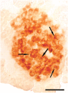
Huntingtin-associated protein 1 (HAP1) was originally found to be localized in neurons and is thought to play an important role in neuronal vesicular trafficking and/or organelle transport. Based on functional similarity between neuron and endocrine cell in vesicular trafficking, we examined the expression and localization of HAP1 in the rat endocrine system using immunohistochemistry. HAP1-immunoreactive cells are widely distributed in the anterior lobe of the pituitary, scattered in the wall of the thyroid follicles, or clustered in the interfollicular space of the thyroid gland, exclusively but diffusely distributed in the medullae of adrenal glands, and selectively located in the pancreas islets. HAP1-containing cells were also found in the mucosa of stomach and small intestine with a distributive pattern similar to that of gastrointestinal endocrine cells. However, no HAP1-immunoreactive cell was found in the cortex of the adrenal gland, the testis, and the ovary. In the posterior lobe of the pituitary, HAP1-immunoreactive products were not detected in the cell bodies but in many stigmoid bodies, one kind of non-membrane-bound cytoplasmic organelle with a central or eccentric electron-lucent core. HAP1-immunoreactive stigmoid bodies were also found in the cytoplasm of endocrine cells in the thyroid gland, the medullae of adrenal gland, the pancreas islets, the stomach, and small intestine. The present study demonstrates that HAP1 is selectively expressed in part of the small peptide-, protein-, and amino-acid analog and derivative-secreting endocrine cells but not in steroid hormone-secreting cells, suggesting that HAP1 is also involved in intracellular trafficking in certain types of endocrine cells.
Figures


Similar articles
-
Selective expression of Huntingtin-associated protein 1 in {beta}-cells of the rat pancreatic islets.J Histochem Cytochem. 2010 Mar;58(3):255-63. doi: 10.1369/jhc.2009.954479. Epub 2009 Nov 9. J Histochem Cytochem. 2010. PMID: 19901268 Free PMC article.
-
Immunohistochemical relationships of huntingtin-associated protein 1 with enteroendocrine cells in the pyloric mucosa of the rat stomach.Acta Histochem. 2020 Dec;122(8):151650. doi: 10.1016/j.acthis.2020.151650. Epub 2020 Nov 6. Acta Histochem. 2020. PMID: 33161374
-
Characterisation of CART-containing neurons and cells in the porcine pancreas, gastro-intestinal tract, adrenal and thyroid glands.BMC Neurosci. 2007 Jul 11;8:51. doi: 10.1186/1471-2202-8-51. BMC Neurosci. 2007. PMID: 17625001 Free PMC article.
-
Endocrine secretory mechanisms. A review.Am J Pathol. 1975 Apr;79(1):170-88. Am J Pathol. 1975. PMID: 164777 Free PMC article. Review. No abstract available.
-
[Physiological and pharmacological actions of prostaglandins and related substances on the endocrine system].Nihon Rinsho. 1985 Mar;43(3):507-13. Nihon Rinsho. 1985. PMID: 3925197 Review. Japanese. No abstract available.
Cited by
-
Alterations of the Sympathoadrenal Axis Related to the Development of Alzheimer's Disease in the 3xTg Mouse Model.Biology (Basel). 2022 Mar 26;11(4):511. doi: 10.3390/biology11040511. Biology (Basel). 2022. PMID: 35453710 Free PMC article.
-
Huntingtin-associated protein 1 interacts with Ahi1 to regulate cerebellar and brainstem development in mice.J Clin Invest. 2008 Aug;118(8):2785-95. doi: 10.1172/JCI35339. J Clin Invest. 2008. PMID: 18636121 Free PMC article.
-
Regulation of L-type Ca2+ Channel Activity and Insulin Secretion by Huntingtin-associated Protein 1.J Biol Chem. 2016 Dec 16;291(51):26352-26363. doi: 10.1074/jbc.M116.727990. Epub 2016 Sep 13. J Biol Chem. 2016. PMID: 27624941 Free PMC article.
-
Selective expression of Huntingtin-associated protein 1 in {beta}-cells of the rat pancreatic islets.J Histochem Cytochem. 2010 Mar;58(3):255-63. doi: 10.1369/jhc.2009.954479. Epub 2009 Nov 9. J Histochem Cytochem. 2010. PMID: 19901268 Free PMC article.
-
Biological functions and potential therapeutic applications of huntingtin-associated protein 1: progress and prospects.Clin Transl Oncol. 2022 Feb;24(2):203-214. doi: 10.1007/s12094-021-02702-w. Epub 2021 Sep 26. Clin Transl Oncol. 2022. PMID: 34564830 Review.
References
-
- Block-Galarza J, Chase KO, Sapp E, Vaughn KT, Vallee RB, Di-Figlia M, Aronin N. (1997) Fast transport and retrograde movement of huntingtin and HAP 1 in axons. Neuroreport 8: 2247–2251 - PubMed
-
- Dragatsis I, Dietrich P, Zeitlin S. (2000) Expression of the Huntingtin-associated protein 1 gene in the developing and adult mouse. Neurosci Lett 282: 37–40 - PubMed
-
- Engelender S, Sharp AH, Colomer V, Tokito MK, Lanahan A, Worley P, Holzbaur EL, et al. (1997) Huntingtin-associated protein 1 (HAP1) interacts with the p150Glued subunit of dynactin. Hum Mol Genet 6: 2205–2212 - PubMed
-
- Fujinaga R, Kawano J, Matsuzaki Y, Kamei K, Yanai A, Sheng Z, Tanaka M, et al. (2004) Neuroanatomical distribution of Huntingtin-associated protein 1-mRNA in the male mouse brain. J Comp Neurol 478: 88–109 - PubMed
Publication types
MeSH terms
Substances
LinkOut - more resources
Full Text Sources
Research Materials
Miscellaneous

