. 2005 Aug 15;170(4):595-605.
doi: 10.1083/jcb.200506051. Epub 2005 Aug 8.

Transcytosis of NgCAM in epithelial cells reflects differential signal recognition on the endocytic and secretory pathways

Affiliations

Transcytosis of NgCAM in epithelial cells reflects differential signal recognition on the endocytic and secretory pathways

Eric Anderson et al. J Cell Biol. .

Abstract

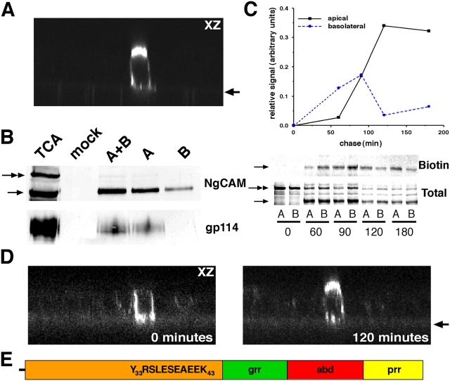
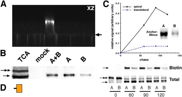
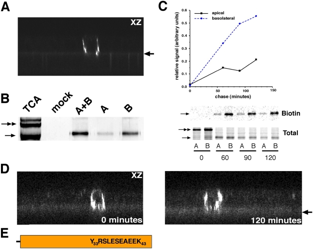
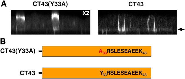
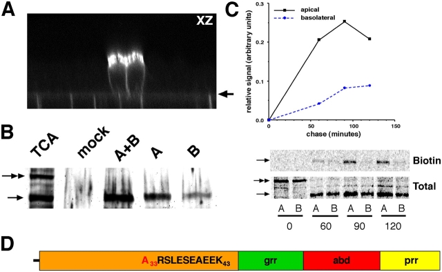
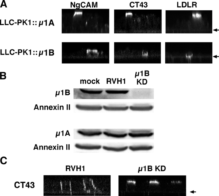
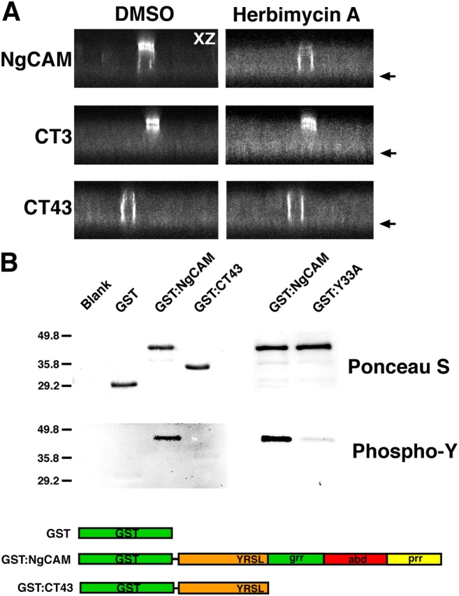
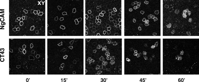
NgCAM is a cell adhesion molecule that is largely axonal in neurons and apical in epithelia. In Madin-Darby canine kidney cells, NgCAM is targeted to the apical surface by transcytosis, being first inserted into the basolateral domain from which it is internalized and transported to the apical domain. Initial basolateral transport is mediated by a sequence motif (Y(33)RSL) decoded by the AP-1B clathrin adaptor complex. This motif is a substrate in vitro for tyrosine phosphorylation by p60src, a modification that disrupts NgCAM's ability to interact with clathrin adaptors. Based on the behavior of various NgCAM mutants, it appears that after arrival at the basolateral surface, the AP-1B interaction site is silenced by phosphorylation of Tyr(33). This slows endocytosis and inhibits basolateral recycling from endosomes, resulting in NgCAM transcytosis due to a cryptic apical targeting signal in its extracellular domain. Thus, transcytosis of NgCAM and perhaps other membrane proteins may reflect the spatial regulation of recognition by adaptors such as AP-1B.


Figures

Figure 1.
Figure 1.
Localization and trafficking of NgCAM. (A) Polarized MDCK cells on Transwell filters were microinjected with NgCAM cDNA and incubated at 37°C for 4 h. The cells were then surface stained with the NgCAM-specific mAb 8D9 and processed for immunofluorescence. The cells were then analyzed by confocal microscopy and a representative x-z section is shown. The arrow denotes the position of the filter. (B) Polarized MDCK cells were infected 16 h with AdNgCAM, placed on ice, and either the apical (A), basolateral, (B) or both (A+B) sides biotinylated. TCA precipitations and mock biotinylations were performed as controls. Biotinylated material was immunoblotted with either pAb G4 (NgCAM) or mAb Y652 (gp114). The 210-kD NgCAM precursor is indicated with a double arrow and the mature 135-kD molecule with a single arrow. Identical results were obtained for the 80-kD fragment, which remains complexed to the 135-kD fragment in a 1:1 ratio (not depicted). (C) Polarized MDCK cells were infected 16 h with AdNgCAM, pulse-labeled with [35S]Met/Cys for 15 min, and chased at 37°C. The cells were then biotinylated on either the apical (A) or basolateral (B) surfaces. NgCAM was immunoprecipitated, and the antibody complexes were brought down with protein G–Sepharose beads. The immunoprecipitated material was boiled off the beads and 20% of the total protein was set aside (Total). The remaining material was applied to NeutrAvidin beads. After binding, the beads were spun down and resuspended in SDS-PAGE sample buffer (Biotin). All samples were then subjected to SDS-PAGE and quantitative autoradiography was performed. The strength of the “Biotin” signal was quantitated and normalized to the “Total” signal, adjusting for the relative inputs. (D) Polarized MDCK cells were infected 16 h with AdNgCAM and conditioned hybridoma supernatant containing mAb 8D9 was added to the basolateral chamber. After 30 min at 37°C, the cells were either fixed (0 min) or growth medium was added and the cells were incubated at 37°C for 120 min and then fixed. The arrow denotes the position of the filter. (E) Schematic of the 114-aa cytoplasmic tail of NgCAM. Note that in all constructs, numbering is from the first residue of the cytoplasmic tail, Lys1.
Figure 2.
Figure 2.
Localization and trafficking of CT3. (A) Polarized MDCK cells were microinjected with CT3 cDNA and processed as described in Fig. 1 A. The arrow denotes the position of the filter. (B) Polarized MDCK cells were infected 16 h with AdCT3 and subjected to vectorial biotinylation as described in Fig. 1 B. (C) Pulse-chase biotinylation analysis of CT3 trafficking was performed as described in Fig. 1 C. (inset) The lumenal domain of NgCAM contains a cryptic apical targeting signal. Polarized MDCK cells grown on 24-mm Transwell filters were infected overnight with AdAnchor-Minus. The medium from either the apical or basolateral chambers was then harvested and NgCAM was immunoprecipitated with mAb 8D9. Western blotting for NgCAM was performed with pAb G4. (D) Schematic of the 3-aa cytoplasmic tail of CT3.
Figure 3.
Figure 3.
Localization and trafficking of CT43. (A) Polarized MDCK cells were microinjected with cDNA encoding CT43 and processed as described in Fig. 1 A. The arrow denotes the position of the filter. (B) Polarized MDCK cells were infected overnight with AdCT43 and subjected to vectorial biotinylation as described in Fig. 1 B. (C) Pulse-chase biotinylation analysis of CT43 trafficking was performed as described in Fig. 1C. (D) Antibody uptake analysis was performed as in Fig. 1 D. The arrow denotes the position of the filter. (E) Schematic of the 43-aa cytoplasmic tail of CT43.
Figure 4.
Figure 4.
NgCAM contains a tyrosine-based basolateral sorting signal. (A) Polarized MDCK cells were microinjected with cDNA encoding either CT43(Y33A) or CT43. The cells were then processed as described in Fig. 1 A. The arrow denotes the position of the filter. (B) Schematics of the CT43(Y33A) and CT43 cytoplasmic tails.
Figure 5.
Figure 5.
Localization and trafficking of NgCAM(Y33A). (A) Polarized MDCK cells were microinjected with the cDNA encoding NgCAM(Y33A) and processed as described in Fig. 1 A. The arrow denotes the position of the filter. (B) Polarized MDCK cells were infected overnight with AdNgCAM(Y33A) and subjected to vectorial biotinylation as described in Fig. 1 B. (C) Pulse-chase biotinylation analysis was performed as described in Fig. 1 C. (D) Schematic of the 114-aa cytoplasmic tail of NgCAM(Y33A).
Figure 6.
Figure 6.
NgCAM interacts with AP-1B. (A) Polarized LLC-PK1::μ1A or LLC-PK1::μ1B cells were infected with either AdNgCAM, AdCT43, or AdLDLR. After overnight incubation at 37°C, the cells were surface stained with mAb 8D9 (NgCAM) or mAb C7 (LDLR) and fixed and processed for immunofluorescence. Confocal microscopy was performed and representative x-z sections are shown. The arrows denote the position of the filters. (B) MDCK cells were either mock infected or infected with RVH1 empty virus or RVH1 encoding an shRNA directed against μ1B. Equal amounts of whole cell lysates were immunoblotted for μ1B, μ1A, or annexin II. (C) Polarized RVH1 empty virus and μ1B knockdown cells were infected with AdCT43. After overnight incubation at 37°C, the cells were surface labeled with mAb 8D9, fixed and processed for immunofluorescence, and analyzed by confocal microscopy. The arrow denotes the position of the filter.
Figure 7.
Figure 7.
The potential role of tyrosine phosphorylation in NgCAM transcytosis. (A) Polarized MDCK cells infected 16 h with either AdNgCAM, AdCT3, or AdCT43 were treated for 5 h with 12.5 μg/ml herbimycin A or DMSO as a control. Representative confocal x-z sections are shown. The arrows denote the position of the filters. (B) The indicated GST fusion proteins were incubated with purified his-p60src in the presence of ATP. After incubation, the samples were subjected to SDS-PAGE and transferred to nitrocellulose, which was Ponceau S stained (top) and then immunostained with an antiphosphotyrosine pAb (bottom). Blank, a control sample containing no added GST protein. Molecular mass markers (labeled in kD) migrated as indicated.
Figure 8.
Figure 8.
Endocytosis of native NgCAM and CT43 from the basolateral surface. MDCK cells infected 16 h with either AdNgCAM or AdCT43 were stained on the basolateral surface for NgCAM with mAb 8D9. The cells were then refed with growth medium and returned to the 37°C incubator for 0, 15, 30, 45, 60, or 90 min, and then processed for immunofluorescence.

Similar articles

Cited by

References

    1. Ang, A.L., T. Taguchi, S. Francis, H. Fölsch, L.J. Murrells, M. Pypaert, G. Warren, and I. Mellman. 2004. Recycling endosomes can serve as intermediates during transport from the Golgi to the plasma membrane of MDCK cells. J. Cell Biol. 167:531–543. - PMC - PubMed
    1. Apodaca, G., L.A. Katz, and K.E. Mostov. 1994. Receptor-mediated transcytosis of IgA in MDCK cells is via apical recycling endosomes. J. Cell Biol. 125:67–86. - PMC - PubMed
    1. Aroeti, B., and K. E. Mostov. 1994. Polarized sorting of the polymeric immunoglobulin receptor in the exocytotic and endocytotic pathways is controlled by the same amino acids. EMBO J. 13:2297–2304. - PMC - PubMed
    1. Aroeti, B., P.A. Kosen, I.D. Kuntz, F.E. Cohen, and K.E. Mostov. 1993. Mutational and secondary structural analysis of the basolateral sorting signal of the polymeric immunoglobulin receptor. J. Cell Biol. 123:1149–1160. - PMC - PubMed
    1. Barroso, M., and E.S. Sztul. 1994. Basolateral to apical transcytosis in polarized cells is indirect and involves BFA and trimeric G protein sensitive passage through the apical endosome. J. Cell Biol. 124:83–100. - PMC - PubMed

Publication types