. 2005 Aug 15;202(4):551-60.
doi: 10.1084/jem.20042463. Epub 2005 Aug 8.

IL-4 receptor expression on CD8+ T cells is required for the development of protective memory responses against liver stages of malaria parasites

Affiliations

IL-4 receptor expression on CD8+ T cells is required for the development of protective memory responses against liver stages of malaria parasites

Alexandre Morrot et al. J Exp Med. .

Abstract

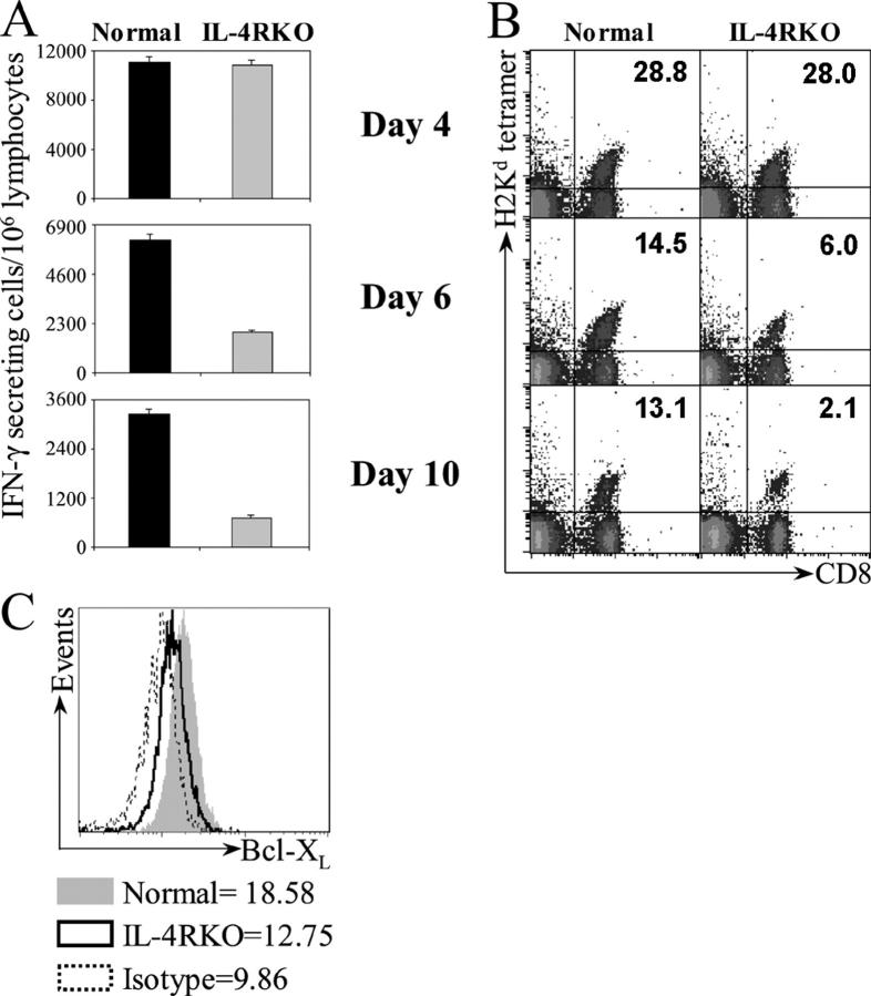
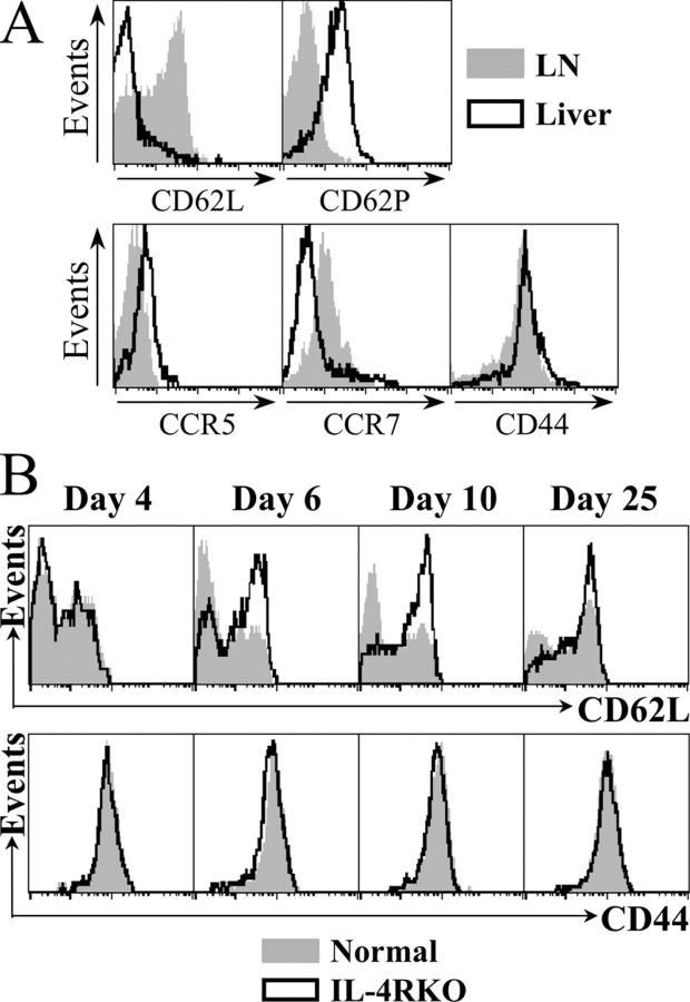
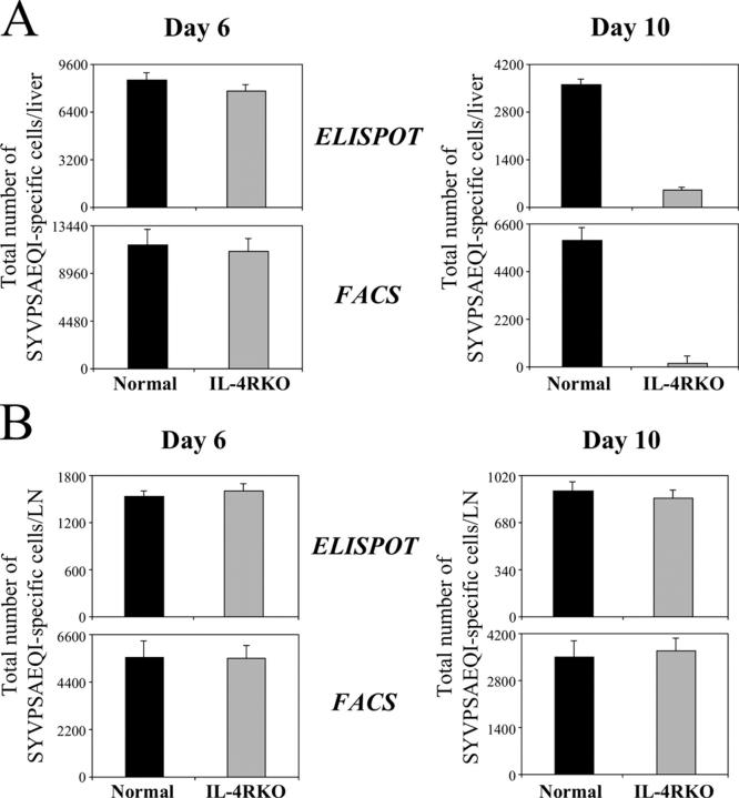
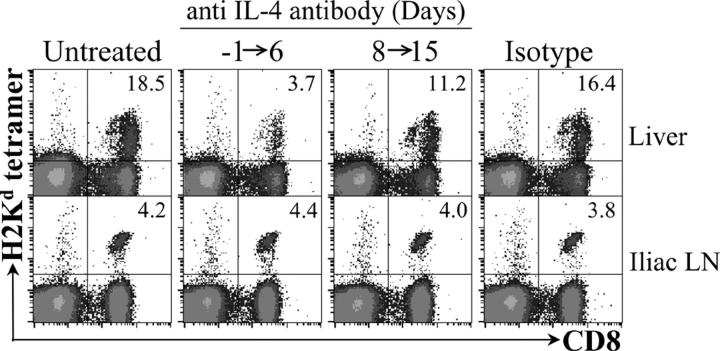
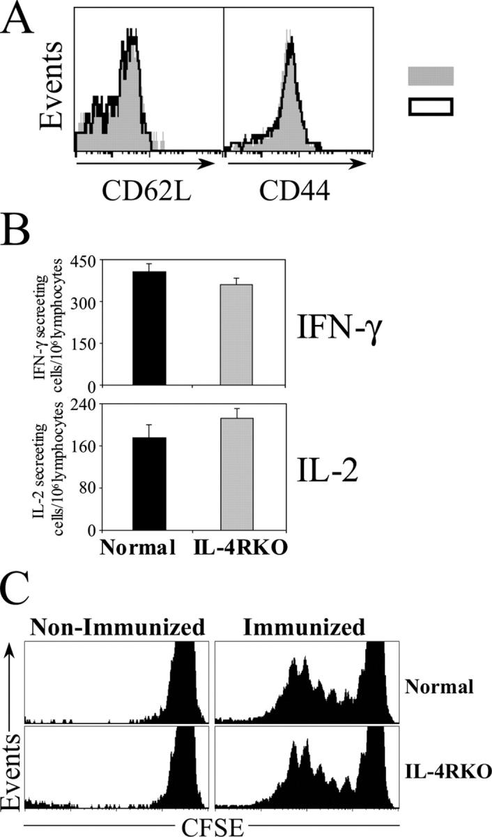
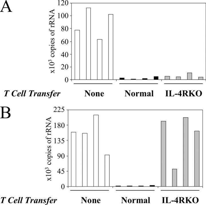
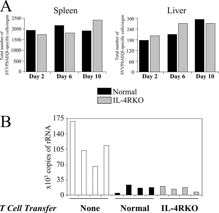
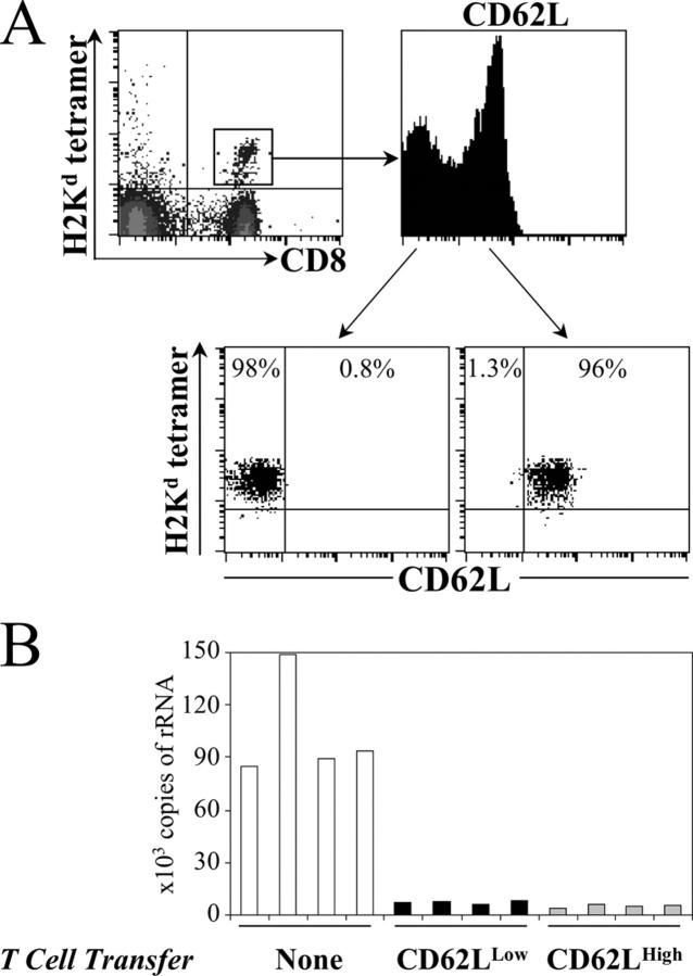
IL-4 receptor (IL-4R)-deficient CD8+ T cells specific for the circumsporozoite protein of Plasmodium yoelii develop a severely impaired memory response after priming with parasites. Memory CD8+ T cells lacking the IL-4R are unable to establish a stable population residing in nonlymphoid organs, although they develop normally in lymphoid organs. Because memory cells from nonlymphoid organs disappear shortly after immunization, the protective antiparasitic activity of this T cell response also is lost. These results demonstrate that IL-4/IL-4R interactions on CD8+ T cells play a critical role in modulating the development and tissue distribution of memory cells induced by parasite immunization. They also indicate that memory cells residing in nonlymphoid tissues are critical for protective immunity against malaria parasites.


Figures

Figure 1.
Figure 1.
IL-4R KO CD8+ T cells develop a reduced response after immunization. Naive normal or IL-4R KO transgenic CD8+ T cells (5 × 10⁵) were transferred into normal BALB/c mice which were subsequently immunized with 5 × 10⁴ irradiation-attenuated sporozoites. The CD8+ T cell response of each was measured in pooled spleen cells, at different days after immunization. Each group consisted of four mice. (A) ELISPOT was used to determine the number of antigen-specific IFN-γ–secreting cells after incubation with antigen (14). Negligible numbers of IFN-γ spots were obtained when cells were incubated without antigen. The data are representative of four independent experiments that yielded the same results. Error bars represent SEM. (B) FACS analysis of this response in spleen was performed by staining with anti-CD8–APC antibodies and tetramer-PE (2). Plots were gated on lymphocytes. The values in the top right corner represent the mean percentages of tetramer+ cells within the total CD8+ T cell population. The data are representative of four independent experiments that yielded the same results. (C) Intracellular Bcl-XL staining of tetramer+/CD8+ T cells at day 4 after immunization. The mean fluorescent intensity of Bcl-XL expression is indicated. Gray line graph identifies normal CD8+ T cells; solid line represents IL-4R KO CD8+ T cells; and dotted line represents Ig isotype control. These data are representative of three independent experiments, using three to five mice per group. The values indicate the mean fluorescent intensity of Bcl-XL expression, the decrease observed in the IL-4R KO cells as compared with the normal cells was statistically significant (P = 0.002; see Fig. S2).
Figure 2.
Figure 2.
Characterization of memory CD8+ T cell subsets and activated IL-4R KO CD8+ T cells. Memory CD8+ T cells were obtained 20 d after immunization from liver and lymph nodes of mice that received normal transgenic CD8+ T cells and were immunized with parasites. (A) Phenotypic analysis of normal memory T cells obtained from liver or lymph nodes (LN) was performed by flow cytometry after staining the cells with tetramer-PE, CD8-APC, and FITC-labeled antibodies against CD62L, CD62P, CCR5, CCR7, and CD44. Line graphs are gated on tetramer+/CD8+ T cells. (B) Expression of CD44 and CD62L in activated CD8+ T cells were assessed using spleen lymphocytes obtained from mice that received normal or IL-4R KO CD8+ T cells, and were immunized with attenuated parasites, as described in the legend of Fig. 1. At specified days after immunization, FACS analysis was performed using anti-CD8–APC antibodies, tetramer-PE, and FITC-labeled antibodies against CD44 or CD62L. The line graphs represent data obtained after gating on tetramer+/CD8+ T cells. Solid gray graphs identify normal CD8+ T cells, and solid lines represent IL-4R KO CD8+ T cells. The increase in the mean fluorescent intensity of CD62L expression observed in the IL-4R KO as compared with the normal cells after day 6 was statistically significant (P < 0.05). These data are representative of four independent experiments, using three mice per group at each time point.
Figure 3.
Figure 3.
IL-4R KO CD8+ T cells do not develop peripheral memory. CD8+ T cell responses developed by normal or IL-4R KO CD8+ T cells were evaluated in different organs and at different time points after immunization with parasites. The adoptive transfer of transgenic CD8+ T cells and immunization with parasites was performed as described in the legend of Fig. 1. Mononuclear cells that were purified from lymphoid and nonlymphoid organs were analyzed by FACS after staining with tetramer-PE and anti-CD8–APC. Plots represent tetramer+/CD8+ lymphocyte pools derived from four mice. The values in the top right corners represent mean percentages of tetramer+ cells within the total CD8+ T cell population. The decrease observed after 10 d in the percentages of IL-4R KO cells from liver, lung, and kidney compared with the normal cells was statistically significant (P < 0.05). These data are representative of four independent experiments, using three mice per group at each time point.
Figure 4.
Figure 4.
Quantification of CD8+ T cell responses in liver and lymph nodes. The total number of SYVPSAEQI–specific T cells present in the liver (A) and lymph nodes (B) of mice was determined 6 and 10 d after adoptive transfer of CD8+ T cells and immunization with 5 × 10⁴ attenuated sporozoites. The estimation of the total number of epitope-specific cells was obtained by multiplying the percentage of tetramer+/CD8+ T cells (FACS) or IFN-γ spots (ELISPOT) by the total numbers of lymphocytes isolated from the respective organs. Each histogram represents data from pooled cells obtained from four mice. These data are representative of four independent experiments, using three mice per group at each time point. Error bars represent SEM.
Figure 5.
Figure 5.
Treatment with anti-IL-4 antibody reduces the number of memory CD8+ T cells in the liver. Naive transgenic CD8+ T cells were transferred into normal mice, some of which received a daily i.p. injection of 0.1 mg of rat antibody against mouse IL-4 (11B11) at each of the indicated days. Control groups received no treatment or a similar amount of nonspecific rat IgG. These mice were immunized with 5 × 10⁴ attenuated sporozoites; 16 d later the number of epitope-specific CD8+ T cells was evaluated by FACS in the liver and iliac lymph nodes of immunized mice. The data of each group were obtained from pooled lymphocytes that were taken from the lymph nodes or the liver. Each group consisted of four mice. Plots were gated on lymphocytes; the number in the upper right corner represents the frequency of tetramer+/CD8+ T cells in the CD8+ population. The data are representative of three independent experiments. The decrease observed in the percentages of the group that was treated with anti–IL-4 antibody during days −1 to 6, as compared with the untreated mice, was statistically significant (P = 0.0196).
Figure 6.
Figure 6.
Phenotypic, functional, and proliferative characteristics of memory CD8+ T cells from lymph nodes. Memory CD8+ T cells were obtained from lymph nodes of mice that received normal or IL-4R KO CD8+ T cells and were immunized with parasites as described in the legend of Fig. 1. 25 d after immunization, these cells were analyzed by FACS. (A) Surface staining with anti-CD8-APC antibodies, tetramer-PE, and CD62L-FITC or CD44-FITC. Solid gray line graphs represent normal CD8+ T cells and solid lines represent IL-4R KO cells. (B) ELIPSOT assay was performed to evaluate the capacity of these cells to produce IL-2 and IFN-γ, after antigen stimulation. Error bars represent SEM. (C) The in vivo proliferation of these memory cells also was assessed using normal and IL-4R KO memory CD8+ T cells obtained from lymph nodes 25 d after immunization. These memory cells were labeled with CFSE, and transferred into mice which were immunized with 5 × 10⁴ irradiation-attenuated sporozoites. The proliferation of the transferred tetramer+/CD8+ memory T cells was evaluated by FACS 3 d after immunization, after staining cells with tetramer and anti-CD8. Line graphs show the CFSE dilution patterns of cells, obtained by FACS after gating on tetramer+/CD8+ T cells. These data are representative of two independent experiments, using three mice per group at each time point.
Figure 7.
Figure 7.
Memory IL-4R KO CD8+ T cells are not protective. An equal number of normal or IL-4R KO transgenic CD8+ T cells were transferred to mice which were immunized 24 h later with 5 × 10³ irradiated sporozoites. These mice were challenged with viable sporozoites at 6 d (A) or 16 d (B) after immunization. 34 h after parasite challenge, the livers of infected mice were excised to measure the parasite load using real-time PCR (20). The endogenous CD8+ and CD4+ T cells were eliminated by treatment with specific antibodies. The histograms represent the number of plasmodial rRNA copies. The data are representative of two independent experiments, using four mice per group at each time point.
Figure 8.
Figure 8.
Homing properties and antiparasitic activity of adoptively transferred memory CD8+ T cells. SIVPSAEQI-specific memory CD8+ T cells were generated from mice that received equal numbers of IL-4R KO naive transgenic cells and were immunized with irradiated sporozoites. 20 d after immunization, tetramer+/CD8+ T cells were obtained from spleen. Equal numbers of normal or IL-4R KO tetramer-specific memory CD8+ T cells were transferred to naive mice. (A) Following adoptive transfer, the number of SIVPSAEQI-specific memory CD8+ T cells in the spleen and liver was measured by ELISPOT at the indicated times. To determine the protective capacity of adoptive transferred memory cells, the naive recipient mice were challenged with 3 × 10⁴ viable sporozoites 10 d after transfer of memory cells. 34 h later, the livers of infected mice were excised to measure the parasite load using real-time PCR (20). (B) The histograms represent the number of plasmodial rRNA copies. These data are representative of two independent experiments, using four mice per group.
Figure 9.
Figure 9.
Antiparasitic activity of CD62Llo and CD62Lhi memory CD8+ T cells. SIVPSAEQI-specific memory CD8+ T cells were generated from mice that received naive normal transgenic cells and were immunized with irradiated sporozoites. (A) 20 d after immunization, tetramer+/CD8+ T cells obtained from spleen were costained with anti-CD62L. Line graph shows CD62L expression of tetramer+/CD8+ T cells. (B) To assess the antiparasitic activity of CD62Llow and CD62Lhigh memory CD8+ T cells, these subpopulations were purified by cell sorting using flow cytometry. Equal numbers (1.5 × 10⁶) of CD62Llo or CD62Lhi tetramer-specific CD8+ T cells were transferred to naive mice. 2 d later, recipient mice were challenged with 3 × 10⁴ viable sporozoites. 34 h after parasite challenge, the livers of infected mice were excised to measure the parasite load using real-time PCR (20). The histograms represent the number of plasmodial rRNA copies. The data are representative of two independent experiments, using four mice per group.

References

    1. Wong, P., and E.G.P. Am. 2003. CD8 T cell responses to infectious pathogens. Annu. Rev. Immunol. 21:29–70. - PubMed
    1. Sano, G., J.C. Hafalla, A. Morrot, R. Abe, J.J. Lafaille, and F. Zavala. 2001. Swift development of protective effector functions in naive CD8(+) T cells against malaria liver stages. J. Exp. Med. 194:173–180. - PMC - PubMed
    1. Mueller, S.N., C.M. Jones, C.M. Smith, W.R. Heath, and F.R. Carbone. 2002. Rapid cytotoxic T lymphocyte activation occurs in the draining lymph nodes after cutaneous herpes simplex virus infection as a result of early antigen presentation and not the presence of virus. J. Exp. Med. 195:651–656. - PMC - PubMed
    1. Kaech, S.M., S. Hemby, E. Kersh, and R. Ahmed. 2002. Molecular and functional profiling of memory CD8 T cell differentiation. Cell. 111:837–851. - PubMed
    1. Lanzavecchia, A., and F. Sallusto. 2002. Progressive differentiation and selection of the fittest in the immune response. Nat. Rev. Immunol. 2:982–987. - PubMed

Publication types