. 2005 Aug 8;33(14):4485-95.
doi: 10.1093/nar/gki756. Print 2005.

Identification and analysis of ribonuclease P and MRP RNA in a broad range of eukaryotes

Affiliations

Identification and analysis of ribonuclease P and MRP RNA in a broad range of eukaryotes

Paul Piccinelli et al. Nucleic Acids Res. .

Abstract

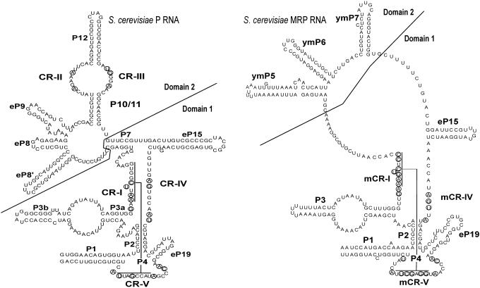
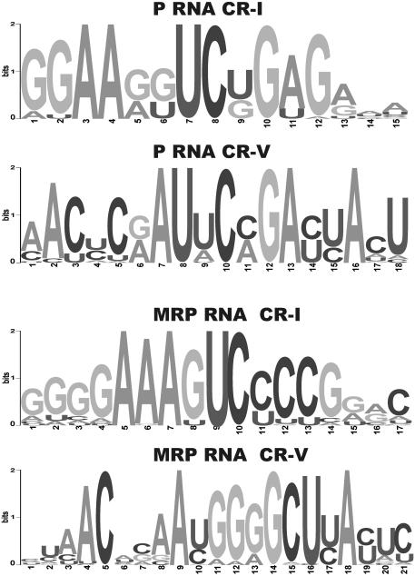
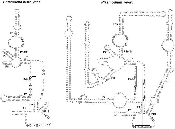
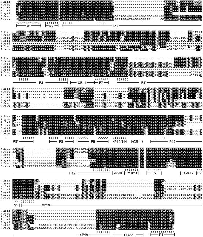
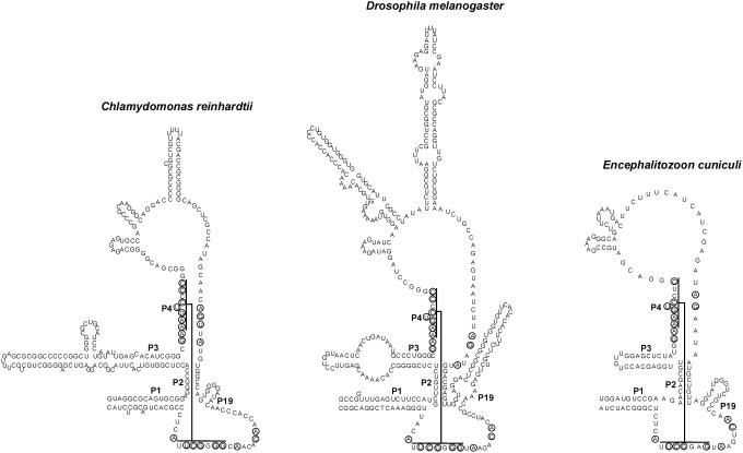
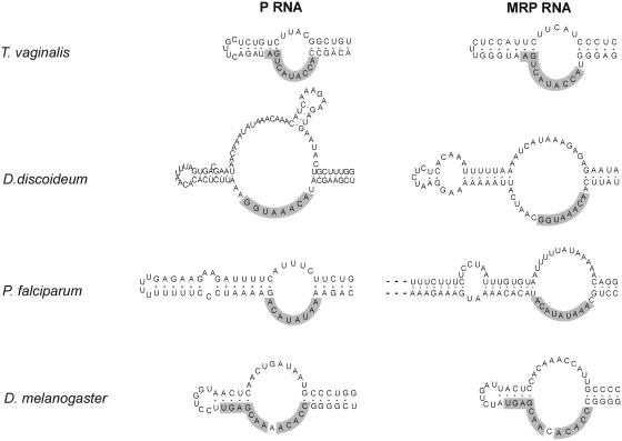
RNases P and MRP are ribonucleoprotein complexes involved in tRNA and rRNA processing, respectively. The RNA subunits of these two enzymes are structurally related to each other and play an essential role in the enzymatic reaction. Both of the RNAs have a highly conserved helical region, P4, which is important in the catalytic reaction. We have used a bioinformatics approach based on conserved elements to computationally analyze available genomic sequences of eukaryotic organisms and have identified a large number of novel nuclear RNase P and MRP RNA genes. For MRP RNA for instance, this investigation increases the number of known sequences by a factor of three. We present secondary structure models of many of the predicted RNAs. Although all sequences are able to fold into the consensus secondary structure of P and MRP RNAs, a striking variation in size is observed, ranging from a Nosema locustae MRP RNA of 160 nt to much larger RNAs, e.g. a Plasmodium knowlesi P RNA of 696 nt. The P and MRP RNA genes appear in tandem in some protists, further emphasizing the close evolutionary relationship of these RNAs.


Figures

Figure 1
Figure 1
Models of S.cerevisiae RNase P and MRP RNAs. Nucleotides that are conserved in all known P and MRP RNAs, respectively, are circled. P RNA is labeled as described previously by Frank et al. (44). CR-I through CR-V are conserved regions. CR-I and CR-V form the helix P4 as indicated. CR-II, CR-III and CR-IV have consensus sequences AGARA, UGNNA and AGNNNNAU, respectively. The nomenclature for the helices P1, P2, P3, P4, P7, P10/11 and P12 is based on homologous bacterial counterparts. In the case of the helices eP8, eP9, eP15 and eP19 the homology to the bacterial counterparts is tentative only. In addition to commonly accepted nomenclature, the helix 5′ of eP8 is here labeled as eP8′. MRP RNA is labeled in a manner similar to P RNA using the nomenclature in Li et al. (15). The structure includes helices ymP5, ymP6 and ymP7 shown to be present in yeast MRP RNA. Conserved regions labeled mCR-I, mCR-IV and mCR-V are counterparts to the CRs in P RNA. mCR-IV has the consensus sequence AGNNA. Figure is in part based on drawings in Walker and Avis (41), although the potential P7 interactions in MRP RNA are not shown here. For details as to CR-I, CR-V, mCR-I and mCR-V see also legend to Figure 2.
Figure 2
Figure 2
Sequence logos of the CR-I and CR-V regions of P and MRP RNA. Logos were created using multiple alignments of available P and MRP sequences and software from Weblogo (27). For multiple alignments see Web Supplement at . P RNA CR-I and CR-V and MRP RNA CR-I and CR-V correspond to positions 1–12, 2–16, 4–13 and 4–22, respectively, in the sequence logos shown.
Figure 3
Figure 3
Secondary structure models of novel eukaryotic P RNAs. P RNAs shown are from Entamoeba histolytica and Plasmodium vivax. The helix numbering is as described previously by Frank et al. (44) and the extra helix present in P.vivax is labeled as P8′. As in Figure 1 interactions of CR-I and CR-V of the P4 helix are shown.
Figure 4
Figure 4
Multiple alignment of Plasmodium P RNAs. Organisms shown are P.berghei, P.yoelii, P.falciparum, P.reichenowi, P.gallinaceum, P.knowlesi and P.vivax. Alignment is based on a ClustalW alignment (45) using gap extension penalty = 0 and was manually edited in the P8′ region to achieve a better structural alignment. Consensus elements are highlighted with BOXSHADE. Positions of helices and conserved regions referred to in Figure 1 are indicated. Angular, curved, square and curly brackets indicate base pairing. Not all paired regions in P3, P8′, P12 and eP19 are shown.
Figure 5
Figure 5
Secondary structure models of selected novel eukaryotic MRP RNAs. RNAs shown are from Chlamydomonas reinhardtii, D.melanogaster and Encephalitozoon cuniculi. Secondary structure of D.melanogaster was inferred from a comparative approach using available Drosophila sequences as described in the text.
Figure 6
Figure 6
Sequence conservation between P and MRP RNA P3 helices. Sequences identical in P and MRP RNA are shaded.

References

    1. Xiao S., Scott F., Fierke C.A., Engelke D.R. Eukaryotic ribonuclease P: a plurality of ribonucleoprotein enzymes. Annu. Rev. Biochem. 2002;71:165–189. - PMC - PubMed
    1. Lygerou Z., Mitchell P., Petfalski E., Seraphin B., Tollervey D. The POP1 gene encodes a protein component common to the RNase MRP and RNase P ribonucleoproteins. Genes Dev. 1994;8:1423–1433. - PubMed
    1. Schmitt M.E., Clayton D.A. Nuclear RNase MRP is required for correct processing of pre-5.8S rRNA in Saccharomyces cerevisiae. Mol. Cell. Biol. 1993;13:7935–7941. - PMC - PubMed
    1. Lygerou Z., Allmang C., Tollervey D., Seraphin B. Accurate processing of a eukaryotic precursor ribosomal RNA by ribonuclease MRP in vitro. Science. 1996;272:268–270. - PubMed
    1. Chu S., Archer R.H., Zengel J.M., Lindahl L. The RNA of RNase MRP is required for normal processing of ribosomal RNA. Proc. Natl Acad. Sci. USA. 1994;91:659–663. - PMC - PubMed

Publication types

MeSH terms