. 2005 Aug 16;102(33):11799-804.
doi: 10.1073/pnas.0408945102. Epub 2005 Aug 8.

Myc interacts genetically with Tip48/Reptin and Tip49/Pontin to control growth and proliferation during Drosophila development

Affiliations

Myc interacts genetically with Tip48/Reptin and Tip49/Pontin to control growth and proliferation during Drosophila development

Paola Bellosta et al. Proc Natl Acad Sci U S A. .

Abstract

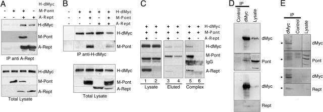
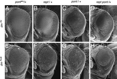
The transcription factor dMyc is the sole Drosophila ortholog of the vertebrate c-myc protooncogenes and a central regulator of growth and cell-cycle progression during normal development. We have investigated the molecular basis of dMyc function by analyzing its interaction with the putative transcriptional cofactors Tip48/Reptin (Rept) and Tip49/Pontin (Pont). We demonstrate that Rept and Pont have conserved their ability to bind to Myc during evolution. All three proteins are required for tissue growth in vivo, because mitotic clones mutant for either dmyc, pont,or rept suffer from cell competition. Most importantly, pont shows a strong dominant genetic interaction with dmyc that is manifested in the duration of development, rates of survival and size of the adult animal and, in particular, of the eye. The molecular basis for these effects may be found in the repression of certain target genes, such as mfas, by dMyc:Pont complexes. These findings indicate that dMyc:Pont complexes play an essential role in the control of cellular growth and proliferation during normal development.


Figures

Fig. 1.
Fig. 1.
Rept and Pont associate with dMyc in vivo. (A and B) M-Pont and/or A-Rept were transiently transfected into S2 cells stably expressing H-dMyc (as indicated). (Upper) Whole-cell lysates were immunoprecipitated with anti-AU1 antibodies for A-Rept (A) or anti-HA antibodies recognizing H-dMyc (B), followed by immunoblotting with anti-tag antibodies to detect the proteins indicated on the right. (Lower) Immunoblots of whole-cell lysates to reveal the relative expression levels of the indicated proteins. Positions of H-dMyc, M-Pont, and A-Rept, respectively, are indicated. The first lane in B Upper contains lysate of nontransfected S2 cells. (C) M-Pont and A-Rept (lanes 1, 3, and 5) or M-Pont alone (lanes 2, 4, and 6) were transiently transfected into S2 cells stably expressing H-dMyc. Cell lysates were incubated with 9E10 antibodies, and the immunoprecipitate was eluted with the 9E10 peptide (lanes 3 and 4). The eluate was then reimmunoprecipitated with anti-AU1 antibodies (lane 5 and 6), and the immunoprecipitate was analyzed by immunoblotting. (D and E) S2 cell lysates (D) or third-instar larval extracts (E) were incubated with anti-dMyc antibodies or control hybridoma supernatant. Immunoprecipitates were blotted with anti-dMyc antibodies, anti-Pont, or anti-Rept antisera as indicated. The rightmost lanes show immunoblots of whole-cell lysates. Asterisks indicate the migration of the endogenous proteins.
Fig. 2.
Fig. 2.
Consequences of pont and/or rept inactivation in vivo. (A) The sizes of pont-/-, rept-/-, or rept-/-pont-/- homozygous mutant larvae are shown in comparison with those of wild-type larvae. Larvae of the indicated genotypes were reared at 25°C and photographed at the indicated times after egg deposition (in days). No rept pont double mutant larvae are alive after day 5. (B and C) Analysis of mitotic pont (B)or rept (C) clones in third-instar imaginal wing discs. Seventy-two hours after their induction, clones of cells with a homozygous mutation for pont (B) or rept (data not shown) are consistently smaller than their corresponding twin clones or even completely absent. (C)If the clones are induced in animals heterozygous for a Minute mutation, their growth is less disadvantaged and their size is still important 120 h after induction in wing imaginal discs, as shown for clones with a homozygous mutant for rept. (B Inset) A pont mutant clone is shown in higher magnification (outlined with white dots in B, the corresponding twin spot is marked by bright color and outlined with a solid line in B Inset). (C) The rept-/- clones are marked by the absence of color; the corresponding twin spots do not survive.
Fig. 3.
Fig. 3.
Heterozygosity for pont or rept results in eye defects in hypomorphic dmyc mutants. Representative scanning electron micrographs of adult eyes. The genotypes shown are dmP0/Y; +/+ (A), dmP0/Y; rept-/+ (B), dmP0/Y; pont-/+ (C), dmP0/Y; rept-/+ pont-/+ (D), ey>dmPL35/Y; ±/+ (E), ey>dmPL35/Y; rept-/+ (F), ey>dmPL35/Y; pont+/+ (G), ey>dmPL35/Y; rept-/+ pont-/+ (H). All pictures are shown at the same magnification; anterior is to the left.

References

    1. Henriksson, M. & Luscher, B. (1996) Adv. Cancer Res. 68, 109-182. - PubMed
    1. Oster, S. K., Ho, C. S., Soucie, E. L. & Penn, L. Z. (2002) Adv. Cancer Res. 84, 81-154. - PubMed
    1. Green, D. R. & Evan, G. I. (2002) Cancer Cell 1, 19-30. - PubMed
    1. Fernandez, P. C., Frank, S. R., Wang, L., Schroeder, M., Liu, S., Greene, J., Cocito, A. & Amati, B. (2003) Genes Dev. 17, 1115-1129. - PMC - PubMed
    1. Zeller, K. I., Jegga, A. G., Aronow, B. J., O'Donnell, K. A. & Dang, C. V. (2003) Genome Biol. 4, R69. - PMC - PubMed

Publication types

MeSH terms