. 2005 Aug 16;102(33):11945-50.
doi: 10.1073/pnas.0505622102. Epub 2005 Aug 8.

Subdomains for transport via plasmodesmata corresponding to the apical-basal axis are established during Arabidopsis embryogenesis

Affiliations

Subdomains for transport via plasmodesmata corresponding to the apical-basal axis are established during Arabidopsis embryogenesis

Insoon Kim et al. Proc Natl Acad Sci U S A. .

Abstract

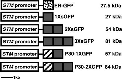
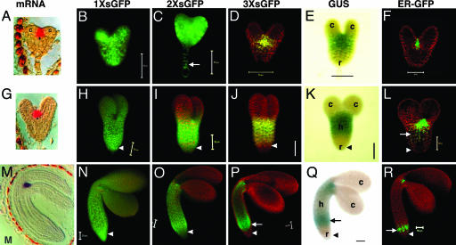
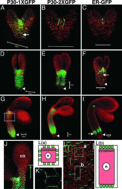
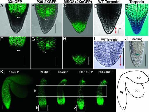
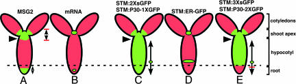
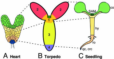
The axial body pattern of Arabidopsis is determined during embryogenesis by auxin signaling and differential gene expression. Here we demonstrate that another pathway, cell-to-cell communication through plasmodesmata (PD), is regulated during apical-basal pattern formation. The SHOOT MERISTEMLESS (STM) promoter was used to drive expression in the shoot apical meristem (SAM) and a subset of cells at the base of the hypocotyl of 1x,2x, and 3x soluble green fluorescent proteins (sGFPs), and the P30 movement protein of Tobacco mosaic virus (TMV) translationally fused to 1x and 2x sGFP. In the early heart stage, 2x sGFP (54 kDa) moves throughout the whole embryo, whereas 3x sGFP (81 kDa) shows more restricted movement. As the embryo develops, PD apertures are down regulated to form local subdomains allowing transport of different sized tracers. For example, movement of 2x sGFP to the cotyledon, and 3x sGFP to root tips, becomes restricted. Subdomains of cell-to-cell transport align with the apical-basal embryo body axis and correspond to the shoot apex, cotyledons, hypocotyl, and root. Studies with P30-GFP fusions reinforce the distinction between embryonic symplastic subdomains. Although P30 targets embryo cell walls as puncta (diagnostic for functional localization of P30 to PD in adult plants), P30 cannot dilate embryonic PD to overcome the barriers for transport between symplastic subdomains, suggesting that specific boundaries separate symplastic subdomains of the embryo. Thus, cell-to-cell communication via plasmodesmata conveys positional information critical to establish the axial body pattern during embryogenesis in Arabidopsis.


Figures

Fig. 1.
Fig. 1.
Constructs transformed into Arabidopsis plants. White box, 3.3-kb STM promoter; dotted box, ER-tethered GFP with signal peptide and KDEL ER retention signal (18); gray box, soluble GFP; striped box, TMV P30.
Fig. 2.
Fig. 2.
Movement pattern of soluble GFP during embryo development. Early heart (AF), late heart (GL), and midtorpedo (MR) embryos for 1× sGFP (B, H, and N), 2× sGFP (C, I, and O), and 3× sGFP (D, J, and P) movement. STM promoter activity is shown by GUS (E, K, and Q) and ER-GFP (F, L, and R). Arrows indicate nucleus in suspensor cells (C), and ectopic expression of STM promoter in hypocotyls (L and PR). Arrowheads indicate root. c, cotyledons; h, hypocotyl; r, root. (Scale bars, 50 μm.) [Images in A, G, and M are reprinted with permission from ref. (Copyright 1998, Company of Biologists, Ltd.).]
Fig. 3.
Fig. 3.
Movement pattern of TMV P30-GFP during embryo development. Early heart (AC), early torpedo (DF), and midtorpedo (GI) embryos for TMV P30–1× GFP (A, D, G, and J), and TMV P30–2× GFP (B, E, and H) movement. ER-GFP shows the site of protein synthesis (C, F, and I). Arrows indicate P30–1× GFP localization to puncta in cell walls (A) and the ectopic expression of STM promoter in hypocotyls (F and I). Arrowheads indicate root. Cells in the box in G are shown in L.(J) P30–1× GFP in the SAM (s) moves several cell layers into cotyledons (co) but stops advancing further. (K) Higher magnification image of P30–1× GFP localizing to puncta in cell walls within SAM. (L) P30–1× GFP targets cell walls, evenly in four sides of cells within (close to) SAM (box a) but preferably goes to apical and basal sides in cells of hypocotyls (box b). Diagrams are individual cells; red represents cytoplasm; green circles represent TMV P30-GFP localized to cell walls as puncta. n, nuclei. (Scale bars, 50 μm.)
Fig. 4.
Fig. 4.
Movement of 3× sGFP and P30–2× GFP in roots of midtorpedo embryos. Overlapping bright field and green GFP fluorescence (AC) and GFP fluorescence alone (FH) are shown. (D and I) Wild-type embryo roots in bright field. (E) Torpedo embryo root showing root cap-specific GUS expression. (J) Seedling root showing quiescent center-specific GUS expression in line QC25 (29). (K) GFP signal in midtorpedo embryos; a, the mid region of hypocotyls; b and c, roots. Arrows indicate cells that mark the boundary between the hypocotyl (hy) and the root (r) in A, B, F, and G. Arrowheads shows nuclear localization of 3× sGFP (A and F). Bidirectional red arrows (D and I) indicate the central root cap. (Scale bars, 50 μm.) Image in I was adapted with permission from ref. . [Image in E reprinted with permission from ref. (Copyright 1999, Company of Biologists, Ltd.).]
Fig. 5.
Fig. 5.
Summary of cell-to-cell transport of symplastic tracers in torpedo embryos. Red represents autofluorescence and green indicates the presence of GFP in cells. Small green circles represent the site of GFP synthesis, and arrows indicate the direction/extent of the cell-to-cell movement of each sGFP from the site of synthesis. Arrowheads mark the boundary between symplastic subdomains of the shoot apex and hypocotyl, and the dotted line shows the boundary between hypocotyls and the root. The MSG2 line expresses 2× sGFP in the SAM and RAM.
Fig. 6.
Fig. 6.
Axial body patterns of Arabidopsis embryos and seedlings. Colors in A and C represent the corresponding clonal regions in early embryos and seedlings. (B) Four subdomains of symplastic transport form along the apical–basal body axis. 1, shoot apex including the meristem (darker green circle) and adjacent cells (orange); 2, cotyledons; 3, hypocotyl; 4, root; co, cotyledons; hy, hypocotyl; ro, root; crc, central root cap; qc, quiescent center. A and C were adapted with permission from ref. .

References

    1. Scheres, B., Wolkenfelt, H., Willemsen, V., Terlouw, M., Lawson, E., Dean, C. & Weisbeek, P. (1994) Development (Cambridge, U.K.) 120, 2475-2487.
    1. Poethig, R., Coe, E. & Johri, M. (1986) Dev. Biol. 117, 392-404.
    1. Saulsberry, A., Martin, P. R., O'Brien, T., Sieburth, L. E. & Pickett, F. B. (2002) Development (Cambridge, U.K.) 129, 3403-3410. - PubMed
    1. Berleth, T. & Chatfield, S. (2002) in The Arabidopsis Book, eds. Somerville, C. & Meyerowitz, E. (Am. Soc. Plant. Biol., Rockville, MD), pp. 1-22.
    1. Laux, T., Wurschum, T. & Breuninger, H. (2004) Plant Cell 16, Suppl., S190-S202. - PMC - PubMed

Substances