Comparative Study
. 2005 Aug 9;65(3):404-11.
doi: 10.1212/01.wnl.0000171450.97464.49.

Increased hippocampal activation in mild cognitive impairment compared to normal aging and AD

Affiliations
Comparative Study

Increased hippocampal activation in mild cognitive impairment compared to normal aging and AD

B C Dickerson et al. Neurology. .

Abstract

Objective: To use fMRI to investigate whether hippocampal and entorhinal activation during learning is altered in the earliest phase of mild cognitive impairment (MCI).

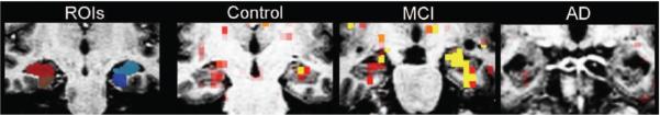
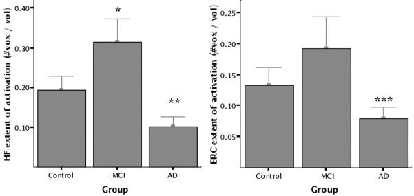
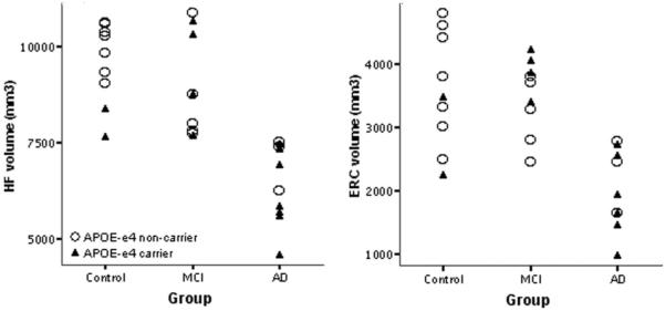
Methods: Three groups of older individuals were studied: 10 cognitively intact controls, 9 individuals at the mild end of the spectrum of MCI, and 10 patients with probable Alzheimer disease (AD). Subjects performed a face-name associative encoding task during fMRI scanning, and were tested for recognition of stimuli afterward. Data were analyzed using a functional-anatomic method in which medial temporal lobe (MTL) regions of interest were identified from each individual's structural MRI, and fMRI activation was quantified within each region.

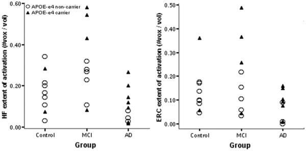
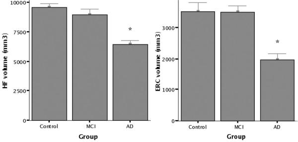
Results: Significantly greater hippocampal activation was present in the MCI group compared to controls; there were no differences between these two groups in hippocampal or entorhinal volumes. In contrast, the AD group showed hippocampal and entorhinal hypoactivation and atrophy in comparison to controls. The subjects with MCI performed similarly to controls on the fMRI recognition memory task; patients with AD exhibited poorer performance. Across all 29 subjects, greater mean entorhinal activation was found in the subgroup of 13 carriers of the APOE epsilon4 allele than in the 16 noncarriers.

Conclusions: The authors hypothesize that there is a phase of increased medial temporal lobe activation early in the course of prodromal Alzheimer disease followed by a subsequent decrease as the disease progresses.


Figures

Figure 1
Figure 1
Representative coronal views of MRI data from single subjects. Regions of interest (ROIs): examples of anatomic ROIs for hippocampal formation (red and light blue) and entorhinal cortex (brown and dark blue), as manually delineated on high resolution T1-weighted structural MRI scans. Control, mild cognitive impairment (MCI), Alzheimer disease (AD): examples of fMRI activation maps from the individual subject in each group with the median extent of HF activation (novel vs repeated contrast, p < 0.01) overlaid on that subject’s T1-weighted structural scan.
Figure 2
Figure 2
Group mean fMRI activation data. Extent of fMRI activation is defined as total (left + right) number of voxels within each region of interest (ROI) showing task-related fMRI activation in novel vs repeated contrast (p < 0.01) divided by total number of voxels in ROI. Greater hippocampal (HF) activation was present in the mild cognitive impairment (MCI) group than controls (*p < 0.03), and lesser hippocampal (**p < 0.005) and entorhinal (ERC; ***p < 0.02) activation was present in the Alzheimer disease (AD) group.
Figure 3
Figure 3
Hippocampal (HF) and entorhinal (ERC) activation by clinical group and APOE genotype. Data points indicate total (left + right) number of voxels within each region of interest (ROI) showing task-related fMRI activation in the novel vs repeated contrast (p < 0.01) divided by total number of voxels in ROI. Among all 29 subjects, greater entorhinal activation was present in APOE E4 carriers than in noncarriers (p < 0.02). MCI = mild cognitive impairment; Alzheimer disease = AD.
Figure 4
Figure 4
Group mean volumetric MRI data. Standard manual volumetric protocols were used to measure regions of interest (ROIs) for the hippocampal formation (HF) and entorhinal cortex (ERC) from each individual subject’s structural MRI. Total volume (left + right) is displayed for each ROI. There was no significant difference in volumes between controls and mild cognitive impairment (MCI), but patients with Alzheimer disease (AD) demonstrated evidence of atrophy (*p < 0.0005).
Figure 5
Figure 5
Hippocampal (HF) and entorhinal (ERC) volume by clinical group and APOE genotype. Data points indicate total (left + right) volume of each region of interest (ROI). As shown in figure 3, the mean volumes of both ROIs are smaller in the Alzheimer disease (AD) group; no APOE effects or group by genotype interactions were observed. MCI = mild cognitive impairment.

References

    1. Small SA, Perera GM, DeLaPaz R, Mayeux R, Stern Y. Differential regional dysfunction of the hippocampal formation among elderly with memory decline and Alzheimer’s disease. Ann Neurol. 1999;45:466–472. - PubMed
    1. Rombouts SA, Barkhof F, Veltman DJ, et al. Functional MR imaging in Alzheimer’s disease during memory encoding. AJNR Am J Neuroradiol. 2000;21:1869–1875. - PMC - PubMed
    1. Kato T, Knopman D, Liu H. Dissociation of regional activation in mild AD during visual encoding: a functional MRI study. Neurology. 2001;57:812–816. - PubMed
    1. Sperling RA, Bates JF, Chua EF, et al. fMRI studies of associative encoding in young and elderly controls and mild Alzheimer’s disease. J Neurol Neurosurg Psychiatry. 2003;74:44–50. - PMC - PubMed
    1. Machulda MM, Ward HA, Borowski B, et al. Comparison of memory fMRI response among normal, MCI, and Alzheimer’s patients. Neurology. 2003;61:500–506. - PMC - PubMed

Publication types

MeSH terms

Substances