Unicameral bone cysts of the pelvis: a study of 16 cases
- PMID: 16089077
- PMCID: PMC1888766
Unicameral bone cysts of the pelvis: a study of 16 cases
Abstract
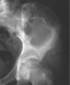
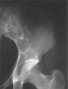
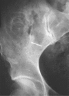
Unicameral bone cysts of the pelvis are extremely rare. This study summarizes the clinical, radiologic and pathologic features of 16 cases. Patients ranged in age from nine to 69. Most lesions were in the anterior portion of the iliac wing; many appeared to be related to an open iliac crest apophysis. This suggests that the pathogenesis of unicameral bone cysts in this portion of the ilium is similar to that seen in the proximal humerus and the proximal femur. The correct diagnosis was made preoperatively in only five cases. This indicates that, although they are well documented, unicameral bone cysts of the pelvis remain a diagnostic problem. Patients received a spectrum of treatments from curettage to observation. There appeared to be no difference in the outcome after any form of treatment. Therefore, unicameral bone cysts of the pelvis can be managed conservatively. The choice to manage patients conservatively depends on making the correct diagnosis based on clinical history and imaging. The most effective imaging is a combination of plain radiographs, computed tomography (CT) and magnetic resonance imaging (MRI).
Conflict of interest statement
Each author certifies that he or she has no commercial associations (e.g. consultancies, stock ownership, equity interest, patent/ licensing arrangements, etc.) that might pose a conflict of interest in connection with the submitted article.
Figures


Similar articles
-
Pathologic fracture through a unicameral bone cyst of the pelvis: CT-guided percutaneous curettage, biopsy, and bone matrix injection.J Vasc Interv Radiol. 2005 Feb;16(2 Pt 1):293-6. doi: 10.1097/01.RVI.0000142598.67437.37. J Vasc Interv Radiol. 2005. PMID: 15713933
-
CT of iliac unicameral bone cysts.AJR Am J Roentgenol. 1981 Jun;136(6):1231-2. doi: 10.2214/ajr.136.6.1231. AJR Am J Roentgenol. 1981. PMID: 6786043 No abstract available.
-
Minimal invasive surgery for unicameral bone cyst using demineralized bone matrix: a case series.BMC Musculoskelet Disord. 2012 Jul 29;13:134. doi: 10.1186/1471-2474-13-134. BMC Musculoskelet Disord. 2012. PMID: 22839754 Free PMC article.
-
Aneurysmal bone cysts of the pelvis in children: a multicenter study and literature review.J Pediatr Orthop. 2005 Jul-Aug;25(4):471-5. doi: 10.1097/01.bpo.0000158002.30800.8f. J Pediatr Orthop. 2005. PMID: 15958897 Review.
-
Unicameral bone cyst of the calcaneus in children.J Pediatr Orthop. 1994 Jan-Feb;14(1):101-4. doi: 10.1097/01241398-199401000-00020. J Pediatr Orthop. 1994. PMID: 8113358 Review.
Cited by
-
Pathological fractures in the paediatric orthopaedic patient population: a current concepts overview of assessment and management.Eur J Orthop Surg Traumatol. 2025 Apr 25;35(1):166. doi: 10.1007/s00590-025-04272-x. Eur J Orthop Surg Traumatol. 2025. PMID: 40274642 Review.
-
A unique case of a unicameral bone cyst in the femoral neck: Successful management in a 14-year-old male.Int J Surg Case Rep. 2025 Jun;131:111356. doi: 10.1016/j.ijscr.2025.111356. Epub 2025 Apr 23. Int J Surg Case Rep. 2025. PMID: 40279993 Free PMC article.
-
Unicameral bone cyst in the pelvis: report of a case treated by placement of screws made from a composite of unsintered hydroxyapatite particles and poly-l-lactide.Rare Tumors. 2019 Dec 12;11:2036361319895075. doi: 10.1177/2036361319895075. eCollection 2019. Rare Tumors. 2019. PMID: 31853343 Free PMC article.
-
Pelvic aneurysmal bone cyst.Biomed Imaging Interv J. 2011 Oct;7(4):e24. doi: 10.2349/biij.7.4.24. Epub 2011 Oct 1. Biomed Imaging Interv J. 2011. PMID: 22279501 Free PMC article.
-
Treatment of unicameral bone cyst: systematic review and meta analysis.J Child Orthop. 2014 Mar;8(2):171-91. doi: 10.1007/s11832-014-0566-3. Epub 2014 Feb 26. J Child Orthop. 2014. PMID: 24570274 Free PMC article.
References
-
- Mirra JM, Bernard GW, Bullough PG, Johnston W, Mink G. Cementum-like bone production in solitary bone cysts (so-called "cementoma" of long bones). Report of three cases. Electron microscopic observations supporting a synovial origin to the simple bone cyst. Clin Orthop. 1978;135:295–307. - PubMed
-
- Virchow R. Ueber die bildung von knochencysten. Monatsber d Kgl Akad D Wissenschaften. 1876. Jun 12, Sitzung der Phisikalischen-mathemat Klasse vom 12 Juni, 1876.
-
- Jaffe H, Lichtenstein L. Solitary unicameral bone cyst. Arch Surg. 1942;44:1004.
-
- Boseker E, Bickel W, Dahlin D. A Clinicopathologic Study of Simple Unicameral Bone Cyst. Surg Gynec and Obstet. 1968;127:550–560. - PubMed
MeSH terms
LinkOut - more resources
Full Text Sources
