

Functional analysis of human hematopoietic stem cell gene expression using zebrafish
- PMID: 16089502
- PMCID: PMC1166352
- DOI: 10.1371/journal.pbio.0030254
Functional analysis of human hematopoietic stem cell gene expression using zebrafish
Abstract
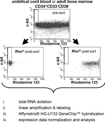
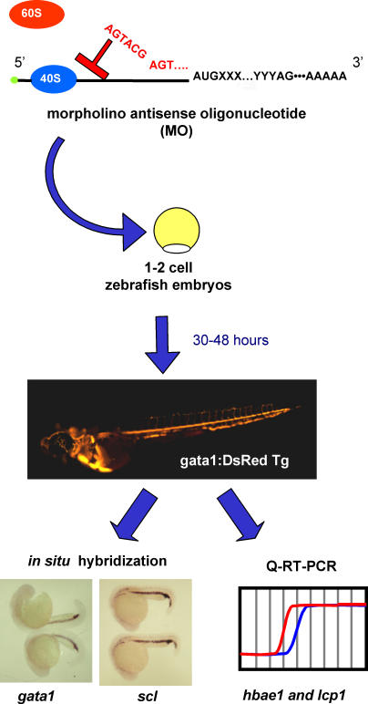
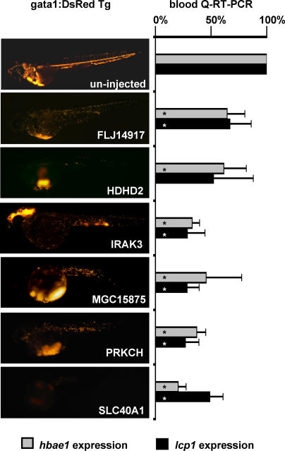
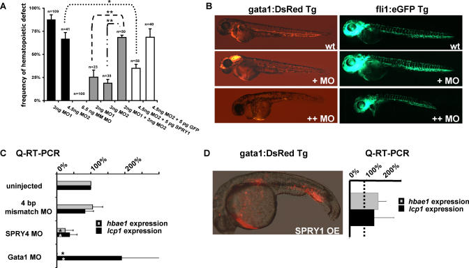
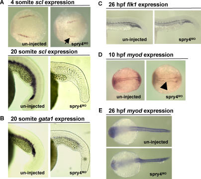
Although several reports have characterized the hematopoietic stem cell (HSC) transcriptome, the roles of HSC-specific genes in hematopoiesis remain elusive. To identify candidate regulators of HSC fate decisions, we compared the transcriptome of human umbilical cord blood and bone marrow (CD34+)(CD33-)(CD38-)Rho(lo)(c-kit+) cells, enriched for hematopoietic stem/progenitor cells with (CD34+)(CD33-)(CD38-)Rho(hi) cells, enriched in committed progenitors. We identified 277 differentially expressed transcripts conserved in these ontogenically distinct cell sources. We next performed a morpholino antisense oligonucleotide (MO)-based functional screen in zebrafish to determine the hematopoietic function of 61 genes that had no previously known function in HSC biology and for which a likely zebrafish ortholog could be identified. MO knock down of 14/61 (23%) of the differentially expressed transcripts resulted in hematopoietic defects in developing zebrafish embryos, as demonstrated by altered levels of circulating blood cells at 30 and 48 h postfertilization and subsequently confirmed by quantitative RT-PCR for erythroid-specific hbae1 and myeloid-specific lcp1 transcripts. Recapitulating the knockdown phenotype using a second MO of independent sequence, absence of the phenotype using a mismatched MO sequence, and rescue of the phenotype by cDNA-based overexpression of the targeted transcript for zebrafish spry4 confirmed the specificity of MO targeting in this system. Further characterization of the spry4-deficient zebrafish embryos demonstrated that hematopoietic defects were not due to more widespread defects in the mesodermal development, and therefore represented primary defects in HSC specification, proliferation, and/or differentiation. Overall, this high-throughput screen for the functional validation of differentially expressed genes using a zebrafish model of hematopoiesis represents a major step toward obtaining meaningful information from global gene profiling of HSCs.
Figures
Similar articles
-
Myeloid-lymphoid initiating cells (ML-IC) are highly enriched in the rhodamine-c-kit(+)CD33(-)CD38(-) fraction of umbilical cord CD34(+) cells.Exp Hematol. 2002 Jun;30(6):582-9. doi: 10.1016/s0301-472x(02)00796-8. Exp Hematol. 2002. PMID: 12063025
-
Microarray and serial analysis of gene expression analyses identify known and novel transcripts overexpressed in hematopoietic stem cells.Cancer Res. 2004 Jul 1;64(13):4434-41. doi: 10.1158/0008-5472.CAN-03-3247. Cancer Res. 2004. PMID: 15231652
-
Characterization of mast cell-committed progenitors present in human umbilical cord blood.Blood. 1999 May 15;93(10):3338-46. Blood. 1999. PMID: 10233886
-
Hematopoietic stem cell development and regulatory signaling in zebrafish.Biochim Biophys Acta. 2013 Feb;1830(2):2370-4. doi: 10.1016/j.bbagen.2012.06.008. Epub 2012 Jun 15. Biochim Biophys Acta. 2013. PMID: 22705943 Review.
-
Assaying hematopoiesis using zebrafish.Blood Cells Mol Dis. 2013 Dec;51(4):271-6. doi: 10.1016/j.bcmd.2013.07.009. Epub 2013 Aug 2. Blood Cells Mol Dis. 2013. PMID: 23916372 Free PMC article. Review.
Cited by
-
Sparc protein is required for normal growth of zebrafish otoliths.J Assoc Res Otolaryngol. 2008 Dec;9(4):436-51. doi: 10.1007/s10162-008-0137-8. Epub 2008 Sep 11. J Assoc Res Otolaryngol. 2008. PMID: 18784957 Free PMC article.
-
Loss of Forkhead box M1 promotes erythropoiesis through increased proliferation of erythroid progenitors.Haematologica. 2017 May;102(5):826-834. doi: 10.3324/haematol.2016.156257. Epub 2017 Feb 2. Haematologica. 2017. PMID: 28154085 Free PMC article.
-
Connective tissue growth factor regulates adipocyte differentiation of mesenchymal stromal cells and facilitates leukemia bone marrow engraftment.Blood. 2013 Jul 18;122(3):357-66. doi: 10.1182/blood-2012-06-437988. Epub 2013 Jun 5. Blood. 2013. PMID: 23741006 Free PMC article.
-
foxm1 Modulates Cell Non-Autonomous Response in Zebrafish Skeletal Muscle Homeostasis.Cells. 2021 May 18;10(5):1241. doi: 10.3390/cells10051241. Cells. 2021. PMID: 34070077 Free PMC article.
-
Meta-analysis of microarray studies reveals a novel hematopoietic progenitor cell signature and demonstrates feasibility of inter-platform data integration.PLoS One. 2008 Aug 13;3(8):e2965. doi: 10.1371/journal.pone.0002965. PLoS One. 2008. PMID: 18698424 Free PMC article.
References
-
- Morrison SJ, Uchida N, Weissman IL. The biology of hematopoietic stem cells. Annu Rev Cell Dev Biol. 1995;11:35–71. - PubMed
-
- Krause DS. Regulation of hematopoietic stem cell fate. Oncogene. 2002;21:3262–3269. - PubMed
-
- Sauvageau G, Thorsteinsdottir U, Eaves CJ, Lawrence HJ, Largman C. Overexpression of HOXB4 in hematopoietic cells causes the selective expansion of more primitive populations in vitro and in vivo. Genes Dev. 1995;9:1753–1765. - PubMed
-
- Antonchuk J, Sauvageau G, Humphries RK. HOXB4 overexpression mediates very rapid stem cell regeneration and competitive hematopoietic repopulation. Exp Hematol. 2001;29:1125–1134. - PubMed
-
- Buske C, Feuring-Buske M, Abramovich C, Spiekermann K, Eaves CJ. Deregulated expression of HOXB4 enhances the primitive growth activity of human hematopoietic cells. Blood. 2002;100:862–868. - PubMed
Publication types
MeSH terms
Substances
Grants and funding
LinkOut - more resources
Full Text Sources
Medical
Molecular Biology Databases
Research Materials
Miscellaneous
