. 2005 Aug;3(8):e257.
doi: 10.1371/journal.pbio.0030257. Epub 2005 Jul 12.

Structure of a pheromone receptor-associated MHC molecule with an open and empty groove

Affiliations

Structure of a pheromone receptor-associated MHC molecule with an open and empty groove

Rich Olson et al. PLoS Biol. 2005 Aug.

Abstract

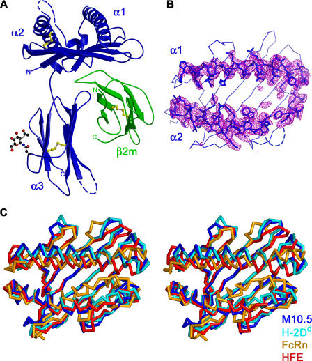
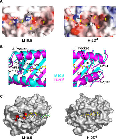
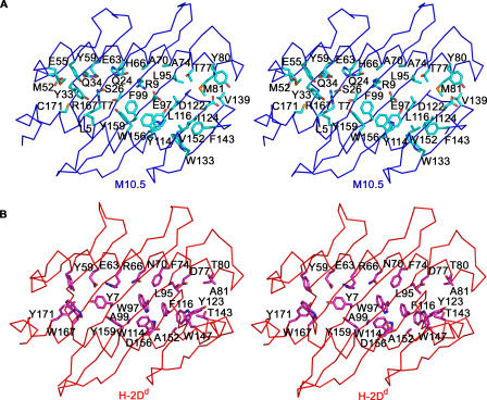
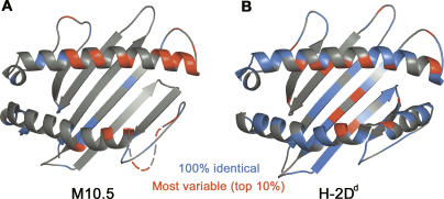
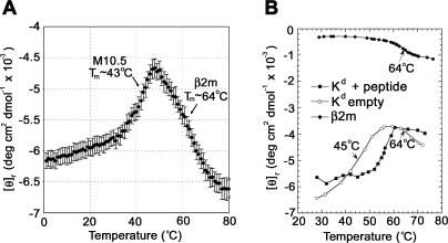
Neurons in the murine vomeronasal organ (VNO) express a family of class Ib major histocompatibility complex (MHC) proteins (M10s) that interact with the V2R class of VNO receptors. This interaction may play a direct role in the detection of pheromonal cues that initiate reproductive and territorial behaviors. The crystal structure of M10.5, an M10 family member, is similar to that of classical MHC molecules. However, the M10.5 counterpart of the MHC peptide-binding groove is open and unoccupied, revealing the first structure of an empty class I MHC molecule. Similar to empty MHC molecules, but unlike peptide-filled MHC proteins and non-peptide-binding MHC homologs, M10.5 is thermally unstable, suggesting that its groove is normally occupied. However, M10.5 does not bind endogenous peptides when expressed in mammalian cells or when offered a mixture of class I-binding peptides. The F pocket side of the M10.5 groove is open, suggesting that ligands larger than 8-10-mer class I-binding peptides could fit by extending out of the groove. Moreover, variable residues point up from the groove helices, rather than toward the groove as in classical MHC structures. These data suggest that M10s are unlikely to provide specific recognition of class I MHC-binding peptides, but are consistent with binding to other ligands, including proteins such as the V2Rs.


Figures

Figure 1
Figure 1. The Structure of M10.5
(A) Ribbon diagram of M10.5 (side view). The heavy chain is blue, the β2m light chain is green, disulfide bonds are yellow, and ordered carbohydrate attached to Asn223 is shown in ball-and-stick representation. Two disordered loops in the heavy chain are shown as dashed blue lines. (B) Top view of the α1–α2 platform overlaid with an Fo-Fc annealed omit electron-density map contoured at 3.5 σ. The map was calculated for one of five molecules in the asymmetric unit using NCS restraints in the annealing process. Residues 55–84 and 137–174, shown in stick representation, were omitted from the structure factor calculation. Electron density is absent for residues 145–150 (dashed line), indicating that they are disordered. No significant electron density is observed in the groove between the α1 and α2 helices. (C) Stereo view of the superposition of the α1–α2 platforms from M10.5, H-2Dd [24], FcRn [32], and HFE [33]. Structures were aligned using residues classified as platform β-sheet residues (see Materials and Methods). The cleft between α1 and α2 helices is significantly narrower in FcRn and HFE than in M10.5 and H-2Dd.
Figure 2
Figure 2. Groove Surface Characteristics
(A) Electrostatic surface representations calculated using GRASP [42] of the M10.5 (left) and H-2Dd (right) grooves. Positions of the six pockets are labeled A–F in yellow. In H-2Dd, Arg62 and Glu163 create a bridge over the peptide-binding groove. In M10.5, Glu63 and Arg167 define a similar feature. The H-2Dd groove is constricted between Asn70 and Arg155. In M10.5, Ala70 and Gly155 lead to a wider groove and continuous D and E pockets. (B) Comparison of residues in the A and F pockets of classical class I MHC molecules and M10.5. All alanine-peptide (yellow, atoms color-coded according to atom type) is derived from the H-2Dd structure [24]. Left: In class I MHC proteins, a cluster of four tyrosine residues in the A pocket (Tyr7, Tyr59, Tyr159, and Tyr171) form hydrogen bonds with backbone atoms in the peptide N-terminus (residues are listed in single letter code for M10.5 and H-2Dd, respectively). In M10.5, Tyr7 is replaced by a threonine residue and Tyr171 is replaced with a cysteine. Five of the six M10 proteins have an additional tyrosine at position 33 that could potentially replace one of the two missing tyrosine residues. M10.5 has a serine instead of a glycine at position 26, which could participate in additional hydrogen-binding interactions. Right: The F pocket of class I MHC molecules is blocked on one end by Thr80, Tyr84, and Lys146. The M10.5 pocket is open on this end (see panels A and C) due to the substitution of a glutamate residue for Tyr84 and the disorder of the loop containing residue 146 (Asp in M10.5). Lys142 in M10.5 is missing sidechain density and has been modeled as an alanine, but most likely would not further occlude the M10.5 F pocket. (C) Molecular surface representations of M10.5 and H-2Dd with an all-alanine peptide (yellow) derived from the H-2Dd structure [24] superimposed in the M10.5 groove. M10.5 atoms that came within 2.5 Å of the peptide trace were considered clashes and are colored red. Four additional alanine residues (green) were added to the C-terminus of the H-2Dd–binding peptide to illustrate that peptides binding in the M10.5 groove could extend out of the F pocket side.
Figure 3
Figure 3. Groove Residues in M10.5 and H-2Dd
Stereo view of residues lining the α1–α2 groove in M10.5 (A) and H-2Dd (B). Groove residues are defined as in Table 2.
Figure 4
Figure 4. Sequence Conservation in M10 and H-2 Loci
(A) Ribbon diagram of the M10.5 α1–α2 platform with positions of residues that are 100% identical in the nine M10 and M1 families colored blue; residues that are among the top 10% most variable are colored red. Variability is determined by the number of amino acids at a given position divided by the frequency of the most common allele at that position. Residues 145–150, which are disordered, are designated by a dashed line. (B) Ribbon diagram of the H-2Dd α1–α2 domain. Conserved (blue) and highly variable (red) residues are derived from an alignment of 19 H-2D alleles [80].
Figure 5
Figure 5. Thermal Denaturation Profiles
Thermal denaturation of M10.5 (A) compared with empty and peptide-filled H-2Kd (B) (modified from Figure 3A in Fahnestock et al. [21]). The CD signal at 223 nm was monitored as a function of temperature for insect cell-derived M10.5 and empty and peptide-filled versions of soluble H-2Kd produced in CHO cells. T ms for the melting of the M10.5 heavy chain and β2m light chain (marked with arrows) were derived by estimating the half-point of the ellipticity change between the beginning and end of each transition. The M10.5 denaturation profile is similar to the profile obtained from empty H-2Kd, suggesting that the M10.5 groove is not occupied. The possibility that polyethylene glycol, a component in the crystallization solutions, might bind to and stabilize M10.5 was investigated by repeating the unfolding experiment in the presence of 20% polyethylene glycol 1000, with no significant changes to the thermal stability profile (data not shown).

Similar articles

Cited by

References

    1. Dulac C, Torello AT. Molecular detection of pheromone signals in mammals: from genes to behaviour. Nat Rev Neurosci. 2003;4:551–562. - PubMed
    1. Leypold BG, Yu CR, Leinders-Zufall T, Kim MM, Zufall F. Altered sexual and social behaviors in trp2 mutant mice. Proc Natl Acad Sci U S A. 2002;99:6376–6381. - PMC - PubMed
    1. Stowers L, Holy TE, Meister M, Dulac C, Koentges G. Loss of sex discrimination and male–male aggression in mice deficient for TRP2. Science. 2002;295:1493–1500. - PubMed
    1. Dulac C, Axel R. A novel family of genes encoding putative pheromone receptors in mammals. Cell. 1995;83:195–206. - PubMed
    1. Matsunami H, Buck LB. A multigene family encoding a diverse array of putative pheromone receptors in mammals. Cell. 1997;90:775–784. - PubMed

Publication types