. 2006 Jan 1;393(Pt 1):43-50.
doi: 10.1042/BJ20051073.

The shed ectodomain of type XIII collagen associates with the fibrillar fibronectin matrix and may interfere with its assembly in vitro

Affiliations

The shed ectodomain of type XIII collagen associates with the fibrillar fibronectin matrix and may interfere with its assembly in vitro

Marja-Riitta Väisänen et al. Biochem J. .

Abstract

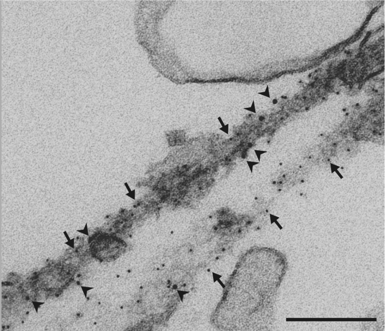
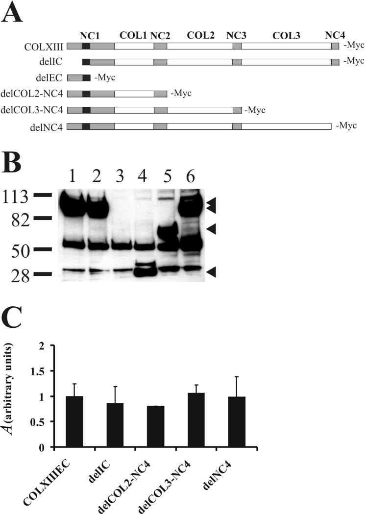
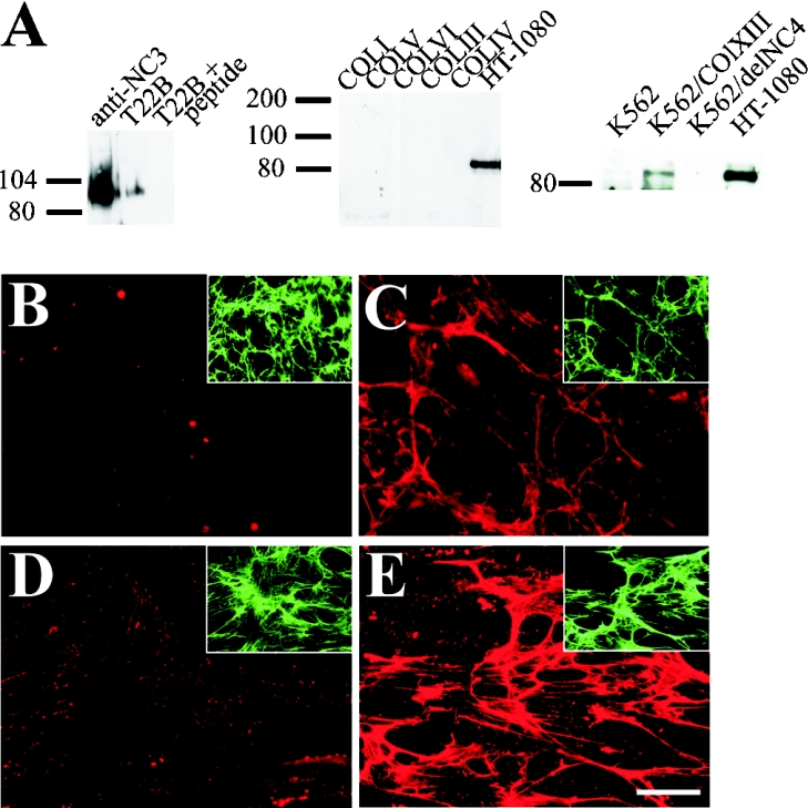
Type XIII collagen is a transmembrane collagen, which is known to exist also as a soluble variant due to ectodomain shedding. Earlier studies with the recombinant ectodomain have shown it to interact in vitro with a number of extracellular matrix proteins, e.g. Fn (fibronectin). In view of its strong binding to Fn, we examined in the present study whether the released soluble ectodomain can bind to the fibrillar Fn matrix under cell-culture conditions and, if so, influence its assembly. In this study, we demonstrate that the type XIII collagen ectodomain of mammalian cells can associate with Fn fibres and may eventually hamper incorporation of the fibrillar Fn meshwork. The association between type XIII collagen and Fn was implicated to be mediated by the C-terminal end of type XIII collagen and the N-terminal end of Fn. The results presented here imply that the shedding of the type XIII collagen ectodomain results in a biologically active molecule capable of remodelling the structure of the pericellular matrix.


Figures

Figure 1
Figure 1. Co-localization of type XIII collagen ectodomain with the Fn matrix
TEM analysis by the immunogold technique showed alignment of the recombinant type XIII collagen ectodomain with fibrillar Fn. Cells were labelled with anti-Fn antibody (shown by arrows) and anti-type XIII collagen NC3 domain antibody (shown by arrowheads) and detected with Protein A conjugated with 5 and 10 nm gold respectively. Scale bar, 200 nm.
Figure 2
Figure 2. Analysis of shedding of type XIII collagen deletion variants
(A) Schematic representation of the full-length type XIII collagen and the deletion variants covering both the ectodomain and the intracellular domains. White boxes denote collagenous sequences, grey boxes non-collagenous sequences and black boxes transmembrane domains. Abbreviations: NC, non-collagenous domain; COL, collagenous domain; IC, intracellular domain; EC, ectodomain. (B) CHO cells were transiently transfected with a given plasmid cDNA for 48 h. The release of type XIII collagen deletion variants into the cell-culture medium was detected by immunoblotting the methanol-precipitated medium with anti-Myc antibody. Sample 1, type XIII collagen ectodomain; sample 2, delIC; sample 3, delEC; sample 4, delCOL2-NC4; sample 5, delCOL3-NC4; sample 6, delNC4. Arrowheads on the right point to the masses of the detected type XIII collagen deletion variants. The 50 and 30 kDa bands in the blot result from unspecific binding of the antibody to unrelated Myc epitopes. The blot shown is representative of three independent assays. (C) Densitometric scanning of the Western blots (n=3) shows that the type XIII collagen ectodomain variants are released from the plasma membranes in comparable amounts with negligible differences. Results are expressed as means with the bars indicating the S.D.
Figure 3
Figure 3. Association of the type XIII collagen ectodomain variants with the fibrillar Fn matrix
CHO cells were transfected with a given plasmid cDNA, with Fn matrix assembly induced by exogenous Fn supplementation at the initiation of transfection. After 48 h, the cells were fixed and stained with anti-Myc and anti-Fn antibodies. Immunofluorescent double stainings showed co-localization of the shed ectodomains of (A) type XIII collagen and (B) delIC with Fn, as shown by the yellow colour in the merged images. (C) delNC4 showed a grossly diminished ectodomain association with Fn, resulting in very faint type XIII collagen and overlay images. In some images, the merged signal at the cell surfaces of the transfected cells is likely to stem from the pericellular Fn fibres overlapping the unshed type XIII collagen present on the plasma membrane. Scale bar, 10 μm.
Figure 4
Figure 4. T22B antibody blocks the type XIII collagen ectodomain association with Fn
(A) T22B detected a protein with identical mass with that recognized by the previously described anti-NC3 domain antibody. Binding was blocked by a peptide corresponding to the epitope of T22B. No cross-reactivity against type I, III, IV, V or VI collagens was observed. HT-1080 cell lysate was included as a positive control. The domain specificity of T22B was verified by the lack of binding to delNC4 variant expressed in K562 cells. In the T22B blocking experiment (B, C) CHO cells with an Fn matrix made of exogenous Fn and (D, E) primary fibroblasts with an endogenously deposited matrix were treated with type XIII collagen ectodomain preincubated with T22B or rabbit non-immune IgG. Immunofluorescent staining with anti-type XIII collagen NC3 domain antibody revealed that the type XIII collagen ectodomain association with the Fn matrix was almost completely blocked with T22B (B, D), but not with non-immune rabbit IgG (C, E). Fn matrices stained with anti-Fn antibody are shown in the insets. Scale bar, 20 μm. Abbreviations: COLI, type I collagen; COLIII, type III collagen; COLIV, type IV collagen; COLV, type V collagen; COLVI, type VI collagen; COLXIII, type XIII collagen.
Figure 5
Figure 5. Binding of Fn70K and Fn45K fragments to type XIII collagen ectodomain
(A) In the preliminary tests with all the Fn fragments, only Fn70K and Fn45K showed binding to the immobilized type XIII collagen ectodomain. (B) Kinetic tests with the Fn70K and Fn45K. The amount of bound analyte was monitored by measuring the variation in the plasmon resonance angle as a function of time and expressed in terms of RU. The recorded RU values, from which the background signal has been subtracted, are shown with various symbols depending on the concentration used. The fitting of the crude data and preparation of the overlay plots were performed using the Biaevaluation 3.0 software. The continuous lines in the graphs indicate the fitted curve. The graphs presented are representative of three individual runs.
Figure 6
Figure 6. Type XIII collagen ectodomain hampers the Fn matrix assembly
Equal numbers of primary fibroblasts were plated and supplemented with exogenous Fn. In (A), the control culture was without additional supplementations, whereas in the others, (B) 50 μg/ml type XIII collagen ectodomain, (C) 100 μg/ml type XIII collagen ectodomain or (D) 50 μg/ml type I collagen was added to the cultures for 3 h. The matrices formed were stained with anti-Fn antibody. (E) The graph shows the quantitative evaluation by image analysis of fluorescence associated with the fibrillar Fn matrices formed, as measured from ten randomly selected viewing fields per sample and expressed in terms of IOD). The sample in (C) was not evaluated quantitatively due to the strong fluorescence associated with the precipitations. Scale bar, 20 μm. (F) Equal numbers of primary fibroblasts were plated in serum-free DMEM and supplemented with type XIII collagen ectodomain, type I or VI collagen for 24 h. The cells were disrupted with 25 mM NH4OH, followed by isolation of the Fn matrices. Equal volumes of samples were separated on an SDS/5% polyacrylamide gel under reducing conditions, followed by Western blotting with anti-Fn antibody. The graph shows results of densitometric scanning of the Western blots, expressed as means with the bars indicating the S.D. The statistical differences were evaluated with Student's t test. *P<0.05, **P<0.01. N.S., not statistical. The blot shown is representative of five independent experiments. Abbreviations: COLXIIIEC, type XIII collagen ectodomain; COLI, type I collagen; COL VI, type VI collagen.

Similar articles

Cited by

References

    1. Geiger B., Bershadsky A., Pankov R., Yamada K. M. Transmembrane extracellular matrix-cytoskeleton crosstalk. Nat. Rev. Mol. Cell Biol. 2001;2:793–805. - PubMed
    1. Vu T. H. Don't mess with the matrix. Nat. Genet. 2001;28:202–203. - PubMed
    1. Behonick D. J., Werb Z. A bit of give and take: the relationship between the extracellular matrix and the developing chondrocyte. Mech. Dev. 2003;120:1327–1336. - PMC - PubMed
    1. Kleinman H. K., Philp D., Hoffman M. P. Role of the extracellular matrix in morphogenesis. Curr. Opin. Biotechnol. 2003;14:526–532. - PubMed
    1. Quaranta V., Giannelli G. Cancer invasion: watch your neighbourhood! Tumori. 2003;89:343–348. - PubMed

Publication types

LinkOut - more resources